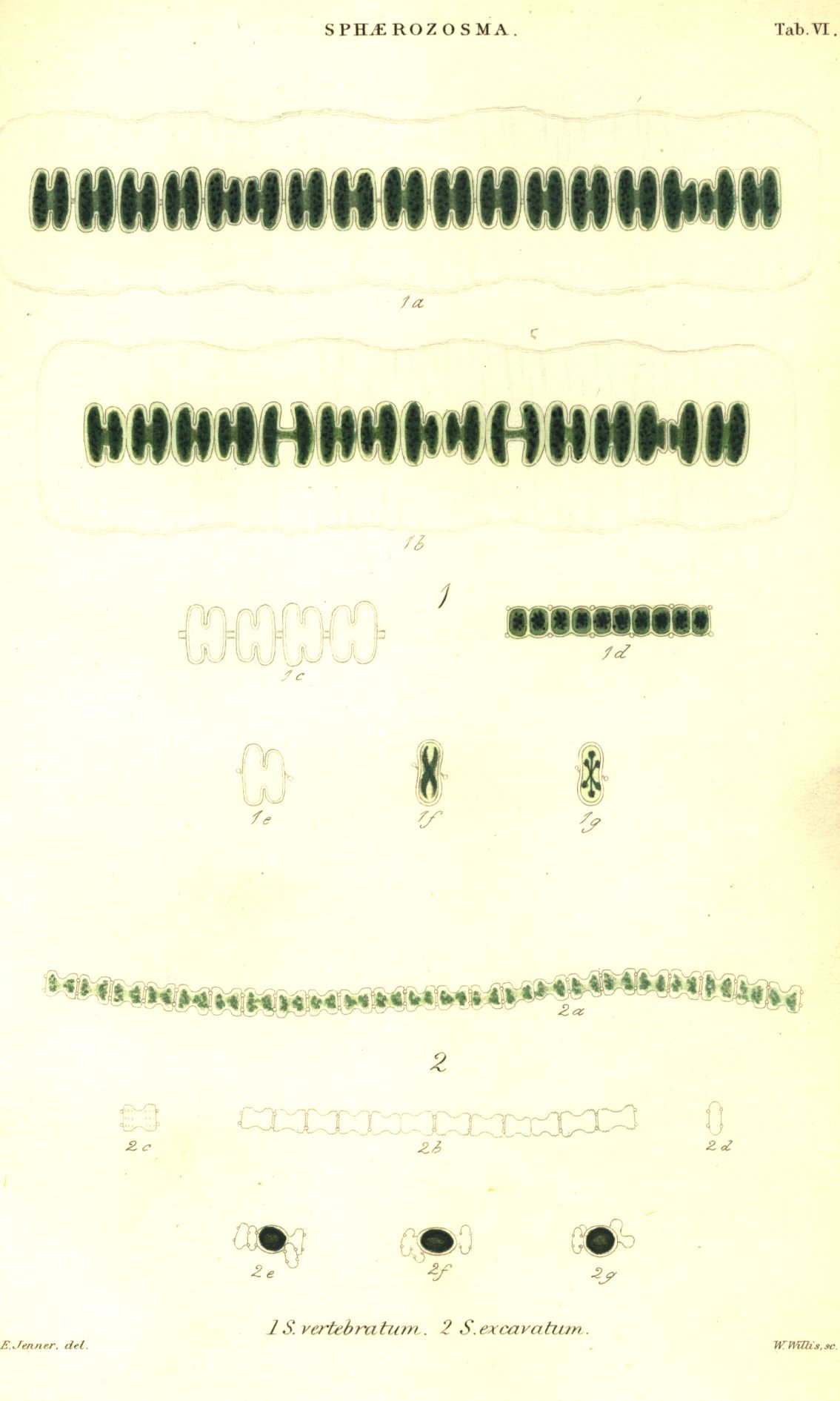
5. SPHAEROZOSMA. Corda
Filament plane, fragile; joints closely united by means of glandular processes, and deeply divided on each side, thus forming two segments and giving a pinnatifid appearance to the filament.
The filaments are pale green, gelatinous, simple, plane, have a pinnatifid appearance from the division of the joints into two segments, are fragile, and finally separate into single joints. I have not observed that the filaments are twisted, as in Desmidium and Didymoprium. At the junction of the joints there are on each margin one or two minute glands or processes which are scarcely discernible in the front view, and do not interfere with the close junction of the joints. The transverse view is linear or oblong, and the processes, one or two at each side, are much more evident than in the front view.
This genus differs from Desmidium, Didymoprium and Hyalothecain its flat filaments (which are not twisted), in the deep division of the joints into segments, and especially in the presence of the minute gland-like processes at the junction of the joints.
On account of its deeply constricted joints, this genus forms a connecting link between the preceding and following genera.
In Sphaerozosma, as in the other genera with deeply constricted cells, the segments are frequently unequal during the growth of the plant, and they become in like manner equal when it approaches maturity and its joints no longer divide.
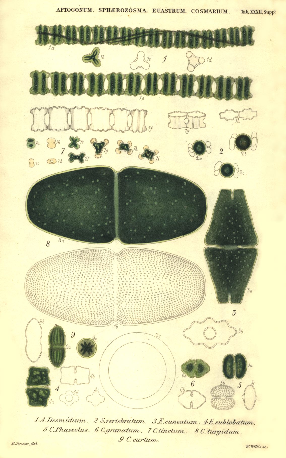
1. S. vertebratum (Bréb.); joints as broad as long, deeply divided into two segments by a narrow notch on each side. Junction-glands oblique, solitary at the centre of each margin.
Desmidium vertebratum, Bréb. Alg. Fal. p. 65. t. 2. (1835).
Hab. Chy-an-hâl and Kerris Moor near Penzance; Dolgelley, J. R. Rotherfield and near Tunbridge Wells, Mr. Jenner. Isle of Sheppey, Mr. Bowerbank. Ambleside, Mr. Sidebotham.
France, De Brébisson. Carlsbad, Corda. North Germany, Ehrenberg and Kützing. West Point, New York, Bailey.
The filaments are much compressed, and the joints, which are nearly equal in length and breadth, are so deeply constricted that at first sight a single one might be mistaken for two. This is more especially the case whilst the endochrome obscures the view of the union of the segments ; as soon however as the joint becomes empty, its nature is distinctly seen. Though in other respects symmetrical, one segment is frequently much smaller than the other.
At the centre, where the joints are connected, is a minute gland or process arising from each margin. The projection of these glands is easily seen, even before the joints separate.
By a transverse view the joints are shown to be compressed, and the oblique glands, which are globular and supported on a very short stipes, are very distinct. In this view the joints are about twice as long as broad, with slightly concave sides, and the endochrome is stellate with from four to six rays.
In a recent state Sphaerozosma vertebratum is very gelatinous and furnished with a broad mucous sheath, which from its tenuity and want of colour is very difficult of detection, and consequently I at first described it as absent. When the plant occurs unmixed with others, the presence of the sheath is indicated by the distinct and parallel filaments exactly as in Hyalotheca mucosa; for in both plants this appearance is produced by its great breadth, which prevents the closer contact of the coloured centres. The sheath is sufficiently apparent in specimens preserved on glass, and is on each side nearly as broad as the filament. In dried specimens it has an irregular waved margin and faint transverse markings.
The greater difficulty of detecting the sheath in recent specimens of Hyalotheca mucosa and S. vertebratum than in Hyalotheca dissiliens and Didymoprium Grevillii, seems to depend more upon its condition in the two former than upon its greater breadth. As in them it is less condensed, it is not only liable to be confounded with the water in which it is viewed, but is also more soluble in it. When Didymoprium Grevillii is mounted in fluid, the sheath remains as distinct as in recent specimens; the case is nearly the same when Hyalotheca dissiliens is so treated, but not so in respect of the present plant, the sheath of' which seems to be dissolved.
Length of joint 1/1429 of an inch; breadth of segments from 1/909 to 1/666; breadth at constriction 1/2237; diameter of sporangium 1/1250.
Table VI. f. 1. a, b. portions of filaments; c. empty joints; d. side view; e. glands in front view; f, g. transverse views.
Tab. VI. f. 2. a, b, c. sporangia.
2. S. excavatum (Ralfs); joints longer than broad, having a deep sinus on both sides, and two sessile glands on each margin at their junction.
Schistochilum excavatum, Ralfs, in Jenner's Fl. of Tunbridge Wells, p. 192 (1845).
Hab. Pools. Dolgelley and Penzance, J. R. Cross-in-hand, and Ashdown Forest, Sussex; bogs at Fisher's Castle, Kent; Farnham, Surrey, and near Southampton, Mr. Jenner. Bristol, Mr. Broome. Amlbleside, Mr. Sidebotham. Rochdale, Mr. Coates.
Falaise, Brébisson. West Point, New York, Bailey.
Very minute, seldom more than twenty-five joints in the filament, which is fragile and finally separates into single joints; at their junction, in the front view, are two minute glands or processes, situated one near each angle, and nearly invisible before the escape of the endochrome. The joints are nearly twice as long as broad and much constricted in the middle; the constriction is like an excavation or broad sinus on each side, so that the margins of the filament appear sinuated. The endochrome is pale bluish green with minute scattered granules.
The transverse view is oblong with four sessile glands, two on each side, and situated near the ends.
From their pale colour and minute size I have experienced much difficulty in determining the form of the angles, which in some specimens seem to me entire, but in others emarginate. Mr. Jenner, using a more powerful microscope, informs me that each is apparently furnished with three minute teeth, which can be detected only when favourably situated for observation. On each segment of the empty joint he finds three transverse series of minute granules, whose appearance at the margins produces this minutely toothed appearance.
I have frequently gathered this species, but always in small quantity, mixed with other Desmidieae ; I am therefore unable to decide whether it possesses a mucous sheath.
The sporangia of S. excavatum have been gathered near Farnham in Surrey, and at Cross-in-hand, Sussex, by Mr. Jenner, who sent his specimen to me for examination. The joints after separation couple; the sporangium is situated between them, and is large compared with the conjugating cells. It is elliptical, and the empty cells are closely connected with it, one at each end. As the specimen was mounted, I was prevented from using the triplet; but from Mr. Jenner's description, the conjugation takes place from their flat or front surfaces, and not in the usual manner from one of the lateral sinuses ; therefore either one of the ends or one of the lateral margins will be presented to the eye.
Length of joints 1/2575 of an inch; breadth of segments 1/3050; breadth at constriction 1/5000; length of sporangium 1/1562; breadth from 1/2325 to 1/1824.
Tab. VI. fig. 2. a. portion of a filament; b. portion of a filament with empty joints; c. empty joint to show the granules; d. transverse view; e, f, g. sporangia.
Sphaerozosma elegans, Corda, Almanach de Carlsbad, 1835, t. 4. fig. 37?; Observ. Micros. sur les Animal. de Carlsbad, p. 21. t. 4. f. 30 ? Hass. Br. Alg. p. 348. t. 84. f. 1.
Odontella ? unidentata, Ehr. Infus. p. 159 (1838).
Isthmia vertebrata, Menegh. Synop. Desmid. in Linnaea 1840, p. 205.
Desmidium compressum, Ralfs, in Annals of Nat. Hist. v. 9. p. 253 (1842).
Sphaerozosma unidentatum, Ralfs, in Annals of Nat. Hist. v. 16. p. 14. t. 3. f. 7. (1845); Trans. of Bot. Society of Edinburgh, v. 2. p. 167.
Isthmosira vertebrata, Kütz. Phycologia Germanica, p. 141 (1845).
Sphaerozosma excavata, Ralfs, in Annals of Nat. Hist. v. 16. p. 15. t. 3. f. 8. (1845); Trans. of Bot. Society of Edinburgh, v. 2. p. 168. Hass. Brit. Freshwater Alg. p. 349.