-   home - index - site map   -   deutsch


Phosphoenolpyruvate:glucose phosphotransferase system (PTS)

from Escherichia coli


To highlight described properties click the boxes . Move the molecule anytime with the mouse.

HPr from Escherichia coli

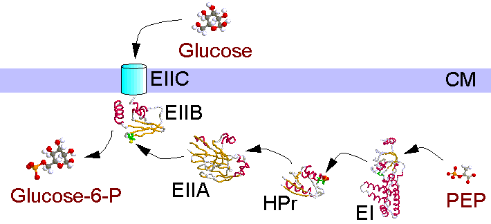
The purpose of the bacterial phosphotransferase system is the specific uptake of sugars into the cells, the sugars are transported uphill a concentration gradient with concomitant phosphorylation. Phosphate donor is the 'energy rich' phosphoenolpyruvate (PEP).The phosphate is transferred via the soluble (and non sugar specific) enzymes EI and HPr to the enzyme complex EII. EII is made up of the components A, B and C, which according to sugar specificity and bacterium involved may be domains of composite proteins. Component/domain C is the permease and anchored to the cytoplasmic membrane. In the glucose PTS of E. coli EIIA is a soluble protein, EIIB/C is membrane bound.

The phosphate group cleaved off the PEP is bound covalently to the proteins to histidines or cysteines. The amount of phosphorylation of the enzymes influences other regulatory mechanisms in the cells (catabolite repression, chemotaxis).


EI EI HPr HPr EIIA EIIA EIIB EIIB


Enzyme I


Enzyme I is phosphorylated by PEP autocatalytically. The whole protein (64 kD) resisted crystallization till now, but the amino terminal part including the active histidine is accessible to structural analysis. The model shown here includes amino acids 2 - 249.

Enzyme I
reset

This part of the protein consists of an alpha-subdomain (amino acids 30-142), and an alpha/beta-subdomain (amino acids 2-19 and 156-229). A four stranded parallel sheet and a three stranded antiparallel sheet form a sandwich with the strands crossed by ~ 90° . One strand is part of both sheets . A helical region is the connection to the (unvisible) carboxy terminal domain.

A comparison of amino acid sequences of enzymes I of different bacteria reveals some amino acids to be conserved completely or conservatively during evolution. This holds especially for the neighborhood of the active histidine . The histidine is phosphorylated at a ring-nitrogen , which in this crystal maintains a hydrogen bridge to an adjacent threonine . In the phosphorylated state the histidine has to occupy a different position, which is sterically possible. The histidine is situated in a groove between the subdomains spacious enough to allow movement of the ring .


HPr


HPr
reset

The histidine containing phosphocarrier protein (HPr) is a molecule of just 85 amino acids. It consists of a four stranded antiparallel sheet , faced by three helices . hprfold Helices a nd c are amphipatic , their hydrophilic parts (blue) face autwards, whereas the hydrophobic amino acids (yellow) are directed to the inside of the protein and together with the pleated sheet form the hydrophobic core of the enzyme. The amino terminus is held in place by an ionic bond between Met1 and Glu70 (located at the amino terminal end of helix c) . NMR spectra of HPr indicate a very strong involvement of the hydroxyl proton of Ser31 in hydrogen bridges, this proton wouldn't exchange with surrounding water . Some loops between the helices and the beta-strands are stabilized by turns (blue).
The amino acid active in phosphate transfer is His15 . After binding of the phosphate to the ring of His15 the protein assumes a different conformation. A superposition of the structures of HPr (white) and P-HPr (blue) indicates the shift in the folding pattern. For some of the side chains sticking out of the protein there are large differences , also His15 shifts its position .

Proteins are not rigid structures, especially side chains may exhibit considerable movability. If protein structures are elucidated by NMR investigations (as in this case) the movability may be demonstrated by simulation of molecular dynamics calculations. In the frame below possible movements in the region around the active histidine (amino acids 14-19) are demonstrated for HPr and P-HPr.


Enzyme II A


EIIAfold

In the phosphorylation chain of the PTS EIIA is the first sugar specifiv enzyme. Its degree of phosphorylation is a sensor for the metabolic state of the cell. Besides transferring the phosphate group from HPr to the permease EIIB/C it also manages chemotaxis toward sugars being transported by the PTS. Additionally it regulates the activity of the adenylate cyclase, of some permeases for non-PTS-sugars and the transcription of some operons. The enzymes molecular weight is 18.1 kD, it consists of 169 amino acids. Atom coordinates for amino acids 19 - 168 are known.

The arrangement of beta-strands in the folding pattern indicates a symmetry of the protein. Within the amino acid sequence there are homologies, by turning the molecule by ~ 180° 46 pairs of Calpha atoms may be superposed with few deviations. Possibly this protein originated by a gene duplication.


Enzyme II A
reset
view down the quasi symmetry axis

The tertiary structure of EIIA is dominated by three pleated sheets . They shape the protein to a distorted barrel . There are few helical stretches . Between Asn32 and Ile45 there is a 310-helix (a), which is connected by a gamma-turn directly to a regular alpha helix (b) (hydrogen bridges shown in green).

EIIA containes two histidines (His75 and His90). His90 is the acceptor for the phosphate group from HPr, His75 is important for the transfer to the permease. Both histidines are situated close together in a groove between helices a/b and c and the rim of the central sheet . They are surrounded by a belt of hydrophobic amino acids (Phe41/71/88, Val39/40/46/96, Ile93) . The amide nitrogen atoms of Asp94 and Thr95 point toward the phosphate, if it is bound to His90 . Asp38/94 which are conserved in EIIAs are important for contacts to other proteins in the reaction chain.


Enzyme II B


The E. coli enzyme II B/C occurs in the membrane as a homodimer. The amino acid chain of domain IIC crosses the membrane eight times; it harbours the sugar binding site. The hydrophilic domain IIB transfers the phosphate group from EIIA to the sugar. The whole protein is built of 477 amino acids. The structure of domain IIB (386-477) was amenable to the determination of its structure.

Enzyme II B
reset

EIIB is a alpha/beta sandwich with a four stranded sheet and three helices to one side . Helix 1 and beta-strands 3 and 4 are hydrophobic (yellow), whrereas helices 2 and 3 as strands 1 and 2 are amphipatic . The hydrophobic core of the domain faces the membrane bound domain C, the amphipatic parts mediate accessibility of components solubilized in the cytoplasm .

The transphosphorylation is performed by a cysteine , which is surrounded by phylogenetically conserved Arg, Asp and Gln . This group of amino acids is framed by hydrophobic amino acids . The somewhat protruding cysteine may react with the recessed phospho histidine in EIIA, the complementary surfaces of the proteins around the reactive centers ease the interaction.


Literature:
PW Postma et al, Phosphoenolpyruvate:carbohydrate phosphotransferase systems of bacteria, Microbiol. Rev. 57 (1993) 543-594
B Erni, Group translocation of glucose and other carbohydrates by the bacterial phosphotransferase system, Int. Rev. Cytol. 137A (1992) 127-148
D-I Liao et al, The first step in sugar transport: crystal structure of the amino terminal domain of enzyme I of the E. coli PEP:sugar phosphotransferase system and a model of the phosphotransfer complex with HPr, Structure 4 (1996) 861-872
NAJ van Nuland et al, The high-resolution structure of the histidine-containing phosphocarrier protein HPr from Escherichia coli determined by restrained molecular dynamics from Nuclear magnetic resonance nuclear Overhauser effect data, J. Mol. Biol. 237 (1994) 544-559
NAJ van Nuland et al, High-resolution structure of the phosphorylated form of the histidine-containing phosphocarrier protein HPr from Escherichia coli determined by restrained molecular dynamics from NMR-NOE data, J. Mol. Biol. 246 (1995) 180-193
D Worthylake et al, Three-dimensional structure of the Escherichia coli phosphocarrier protein IIIglc, Proc. Natl. Acad. Sci. USA 88 (1991) 10382-10386
M Eberstadt et al, Solution structure of the IIB domain of the glucose transporter of Escherichia coli, Biochemistry 35 (1996) 11286-11292
G Gemmecker et al, Glucose transporter of Escherichia coli: NMR characterization of the phosphocysteine form of the IIBGlc domain and its binding interface with the IIAGlc subunit, Biochemistry 36 (1997) 7408-7417


6-99 © Rolf Bergmann   -   http://www.biologie.uni-hamburg.de/lehre/bza/kanal/transp/pts/epts.htm