Click on title or image to view video
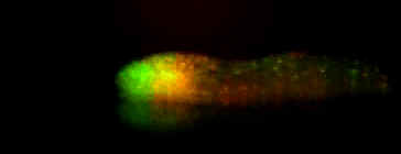 |
Movement of individual prestalk A (ecmA promoter tagged to wild type GFP) and prestalk O (ecmO promoter tagged to redshifted GFP) cells in a D. discoideum slug. Images were captured every 20 seconds. From D. Dormann, T. Abe, J. Williams, and C. Weijer, University of Dundee. |
Prestalk cells during culmination
 |
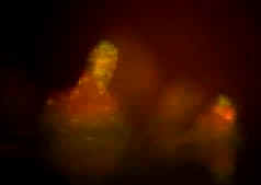
Movement of individual prestalk A (ecmA promoter tagged to wild type GFP) and prestalk O (ecmO promoter tagged to redshifted GFP) cells during culmination. Images were captured every 20 seconds. From D. Dormann, T. Abe, J. Williams, and C. Weijer, University of Dundee. |
Prestalk cells upon slug dissociation
 |
Movement of individual prestalk A (ecmA promoter tagged to wild type GFP) and prestalk O (ecmO promoter tagged to redshifted GFP) cells upon dissociation from the slug. Images were captured every 20 seconds. From D. Dormann, T. Abe, J. Williams, and C. Weijer, University of Dundee. |
 |
Core of a spiral wave in aggregating D. discoideum cells. Time between images is 10 seconds. From F. Siegert and C. J. Weijer, J. Cell Sci. 93, 325-335 (1989). |
 |
Multiple pacemaker centers within a single D. discoideum mound. Time between successive images is 30 seconds. From F. Siegert and C. J. Weijer, Current Biology 5, 937-943 (1995). |
Dark field waves in early aggregation
 |
Dark field waves of D. discoideum cells on caffeine agar. Time between images is 36 seconds. From F. Siegert and C. J. Weijer, J. Cell Sci. 93, 325-335 (1989). |