Prior Page |
 Next Page |
Prior Page |
Next Page |
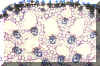
Unlike roots which have a solid, central cylinder of procambium, the procambium in stems occurs in isolated, small cylinders which are distributed in a regular pattern amid the ground tissue. The innermost ground tissue is called the Pith. The external ground tissue is called the Cortex. The stem resembles a column of reinforced concrete. The Epidermis would correspond to the outer mold. The vascular bundles would be steel rods, and the ground tissues would be the concrete that fills the empty volume inside the mold. The distribution of vascular bundles is controlled by the developing leaves and even the youngest primordia may have Procambium located immediately beneath it. The Procambium cells stain densely in the images below.
|
| Long Section of a Shoot Tip showing the distribution of Procambium amid the Ground Tissue of the Pith (Center) and Cortex (Peripheral) |
The Vascular Bundles are called fascicles. This term refers to a Roman symbol which had an axe surrounded by a bundle of sticks, lashed together. The region of ground tissue between the fascicles is called interfascicular. This is best seen in a cross-section of a typical dicot. We will use Coleus to illustrate many aspects of stem anatomy.
|
Coleus stem X-S stained with Phloroglucinol |
Large Vascular Bundles are located in each corner of the Coleus stem. Smaller bundles occur in-between. Thus, they form a single outline in the stem and they define a central Pith and a peripheral Cortex. The overall distribution of fascicles in Coleus is typical for Dicots.The Vascular Bundles are composed of Xylem and Phloem and are called collateral bundles. Fibers may or may not be associated with the vascular bundle.
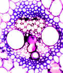
Large Vascular
Bundle from Coleus, stained with Phloroglucinol - Note the Tissues Involved -
Phloroglucinol stains Lignin Red |
|
|
| Same as above but stained with Toluidine Blue - Phloem was not stained in either specimen. |

We will also study Aristolochia stem in detail. We have seen this material previously when we studied Phloem and Xylem. This is a dicot stem and has one ring of vascular bundles in cross-section.
Cross-Section of Aristolochia
Stem |
|
|
|
|
|
|
|
Vascular Tissues in Aristolochia Vascular Bundle |
Phloem in Aristolochia Vascular Bundle |
|
| Commercial
Slide Showing the Vascular Bundles in a Typical Dicot Stem |
|
| Diagram
Showing the Development of Primary Tissues in a Dicot Stem |