|
||
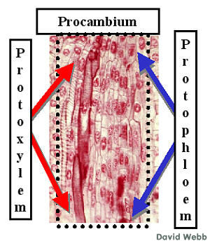 |
Phloem also comes from the Procambium. Protophloem differentiates from the outermost Procambium. Phloem differentiation is harder to follow because the Sieve Elements do not have dramatic cytological features like those in Xylem. | |
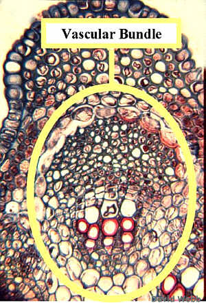 |
Xylem is clearly visible in the attached illustration. It is harder to specifically identify the Phloem. However, careful inspection leads to the conclusion that the tissue immediately opposite the xylem arose from the same shaft of cells (Procambium) as the xylem. Consequently, we can be fairly confident that the nonlignified cells with small diameters, adjacent to the xylem in this vascular bundle are the Phloem. Sometimes it is difficult to specifically point out Phloem based on cytology. Association with xylem is one way that phloem can be identified if only tentatively.Locate the Phloem in this image! | |
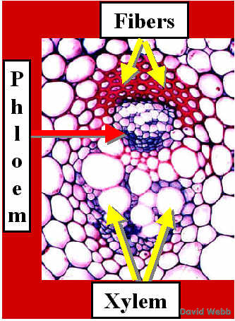 |
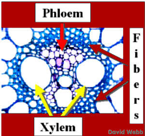
Many plants produce Vascular Bundles which contain a "cap" of Fibers. These are usually associated with the Phloem. Thus, the Phloem can be located between the Fibers and the Xylem. Furthermore, Phloem cells may have a characteristic organizational pattern and cell sizes which help to identify them. | |
 Sugarcane Vascular Bundle stained with Toluidine Blue |
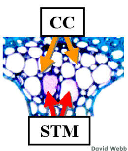
In many monocots like Sugarcane the Phloem is highly organized and has a regular pattern of narrow and wide cells. The wide cells are the Sieve Tube Members. The small cells are Companion Cells. Fibers surround the vascular bundle in this case. | |
 Phloem of a Sugarcane Vascular Bundle stained with Toluidine Blue |
Companion Cells (CC) and Sieve Tube Members (STM) develop from the same progenitor cell. The STM are analogous to Vessel Members. They are columnar cells which unite vertically to form a Sieve Tube. STM are enuclate at maturity and depend on the Companion Cells to regulate physiological processes in the STM. Each STM has one to several CC. The pink color in one of the STM is due to the presence of a Sieve Plate which is analogous to a Perforation Plate. | |
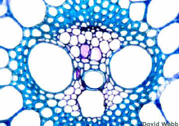 |
Locate the various cells in this unlabeled image of a Sugarcane Vascular Bundle stained with Toluidine Blue. | |
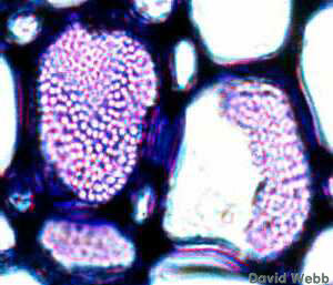 Sieve Plates from the image above. |
Sieve Plates occur on endwalls. of STM. They contain large Sieve Pores. These are much larger than Plasmodesmata. The Plasmalemma and Cytoplasm are continuous from one STM to another through the Sieve Pores. | |
 Lecture Directory |
 Home Page |
Next Page |
|---|---|---|