Stems I
Stems
vary in structure and may be classified into types such 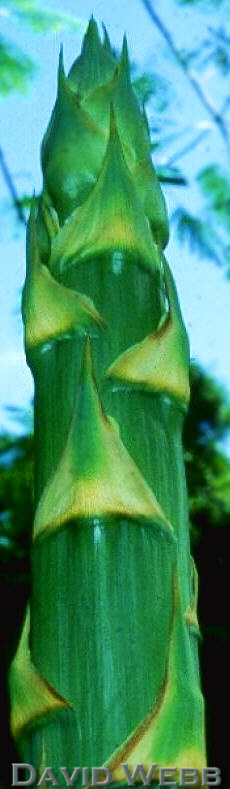 as woody vs. herbaceous. This
classification is artificial, because there is no sharp line of demarcation between these
two, and sometimes the differences are of a purely quantitative kind. The chief
differences between stems are based on the spatial relationship between the vascular
and nonvascular tissues, and by the relative amounts of secondary growth.
as woody vs. herbaceous. This
classification is artificial, because there is no sharp line of demarcation between these
two, and sometimes the differences are of a purely quantitative kind. The chief
differences between stems are based on the spatial relationship between the vascular
and nonvascular tissues, and by the relative amounts of secondary growth.
Stems are more complex in structure than roots mainly because of the complexity of the primary vascular system. This is due to the fact that several vascular bundles may diverge into the leaves. These bundles which are outside of the central ring of vascular bundles are called leaf traces. The size and complexity of leaves is correlated with the complexity of the vascular system in the stem. This lab will consider stems which trace their origin directly to the apical meristem. This is known as primary growth. Some plants stop growing at this point but others many continue to grow due to the activity of lateral meristems. This type of growth is called secondary.
During secondary growth the resemblance between roots and stems increases as the secondary xylem and phloem of roots and stems show many similarities.
Apical Meristems of Vegetative Shoots
Gross characteristics of shoot tips. We can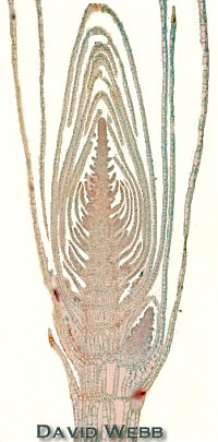 study fresh preparations of shoot tips from Elodea, Artocarpus
(breadfruit), Ficus, Apium (celery) or Coleus
species.
study fresh preparations of shoot tips from Elodea, Artocarpus
(breadfruit), Ficus, Apium (celery) or Coleus
species.
Dissect the shoot tip of Elodea under a stereo scope.
Take a vigorous stem and remove a terminal stem of approximately 4-cm.
Remove the leaves from the basal 2-3 cm..
Blot this dry and embed it in a piece of modeling clay.
Attach
the clay to a plastic Petri plate base so that the stem is erect, and place this on the
stage of a dissecting scope.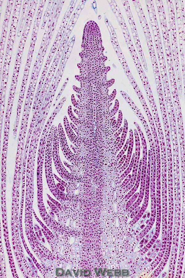
Start removing the most apical leaves carefully with forceps.
The leaves should get smaller and smaller as you approach the shoot tip.
Increase your magnification as you proceed.
You may want to switch to a dissecting needle to remove the smallest leaves by applying gentle pressure.
You
should eventually notice a smooth, shiny, rounded apical meristem with tiny leaf
primordia.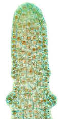
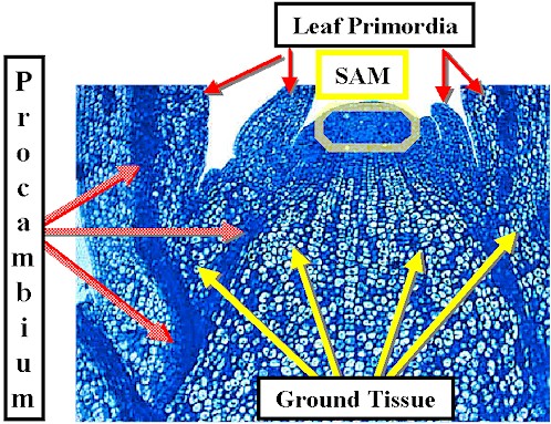
This reveals the uppermost region of tissue initiation called the apical meristem and the subtending zone of leaf initiation.
These are collectively known as the shoot tip. The uppermost leaves are called primordia because they do not possess leaf-like morphology. More well developed leaves can be seen in the basipetal direction (basipetal = away from & towards the base).
Depending on the species, the primordia are arranged spirally, in pairs or in whorls. They appear close to one another because the internodes are not yet extended in the youngest portion of the shoot.
Examine a commercial slide of Elodea and identify the structures cited above.
Commercial longitudinal sections of shoot tips illustrate the beginning of tissue differentiation beneath the apical meristem.
The following tissues and structures may be distinguished:
1. apical meristem;
2. leaf primordia; 3. protoderm;
4. procambium; 5. ground (rib) meristem;
6. axillary buds
Observe the size increases of successively older cells.
Note differences between cells of the procambium and ground meristem in terms of shape, size, and contents.
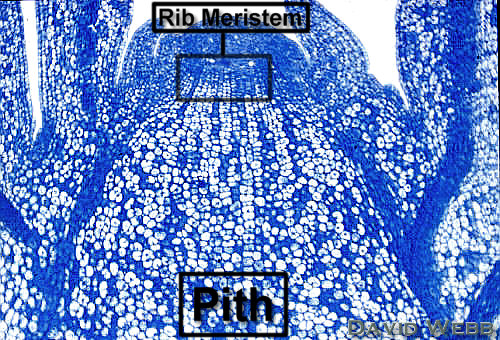
Also find the rib-meristem which produces the pith.
The term Rib Meristem is applied to Ground Meristem which produces the Pith. It is characterized by transverse divisions which produce very orderly cell files. The Ground Meristem that produces the Cortex is not always easy to pinpoint. In my mind both are Ground Meristem but you should understand the term Rib Meristem.
Species with opposite leaves, such as Coleus are particularly convenient for the study of apical differentiation.
It is not possible to see all of these features in one shoot tip. Examine the illustrations below to help you identify the major regions and structures in the shoot tips we have displayed in the lab.
|
|
| Shoot Tip with prominent Rib Meristem | |
Apical Meristems with Apical Cells
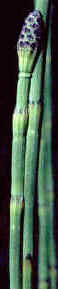
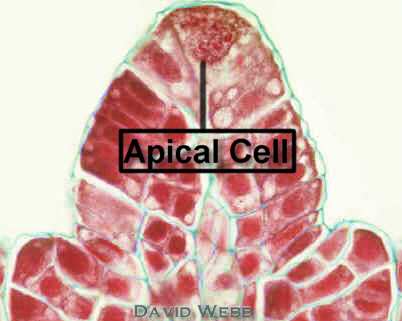
Apical meristem with a single Apical Cell: Equisetum (horsetail) Seed plants have multicellular apical meristems. However, non-seed plants like ferns can have a large Apical Cell which acts as the source of all cells.
Equisetum |
|
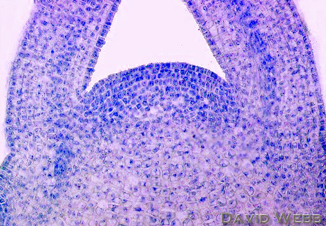
Apical Meristems with Tunica-Corpus Organization.
The tunica,
is one or more superficial layers that show only anticlinal (perpendicular
to the surface) divisions. 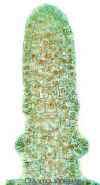
The corpus is a group of cells covered by the tunica. The corpus is characterized by divisions in many planes. This adds volume to the stem as its derivatives enlarge.
The actual number of tunica layers is not always clear. It will be sufficient to discern meristems with One or Several tunica layers.
The corpus can also be hard to delimit. However, when there is a prominent rib meristem, the corpus is circumscribed.
Ricinus
Forsythia
|
|
Forsythia SAM |
SAM with Three Tunica Layers |
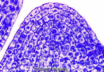
Apical Meristems with Cytohistological Zonation.
The terms cytohistochemical zonation mean that different groups of cells (cyto) respond in a distinct way to various stains (histochemical) .
Gymnosperms
like Norfolk Island Pine (Araucaria) 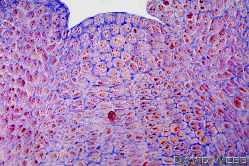 best
exemplify this type of apical meristem. The zonation seen in some plants is due to
differences in the staining density of the cells and in perceived patterns of cell
division. There are a large number of terms which are used to describe different zones in
these meristems. The basic feature of these meristems is the presence of central
mother cells. These are centrally located at the summit of the SAM, and include parts
of the tunica and corpus. The cells are larger and more vacuolate than the surrounding
meristematic cells. Consequently, they stain lightly compared to their neighbors. Finally,
they seem to divide infrequently. The overall organization has a tunica and corpus.
best
exemplify this type of apical meristem. The zonation seen in some plants is due to
differences in the staining density of the cells and in perceived patterns of cell
division. There are a large number of terms which are used to describe different zones in
these meristems. The basic feature of these meristems is the presence of central
mother cells. These are centrally located at the summit of the SAM, and include parts
of the tunica and corpus. The cells are larger and more vacuolate than the surrounding
meristematic cells. Consequently, they stain lightly compared to their neighbors. Finally,
they seem to divide infrequently. The overall organization has a tunica and corpus.
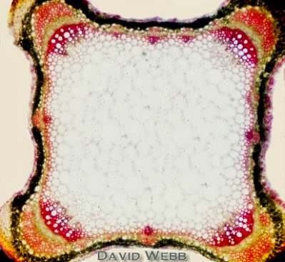
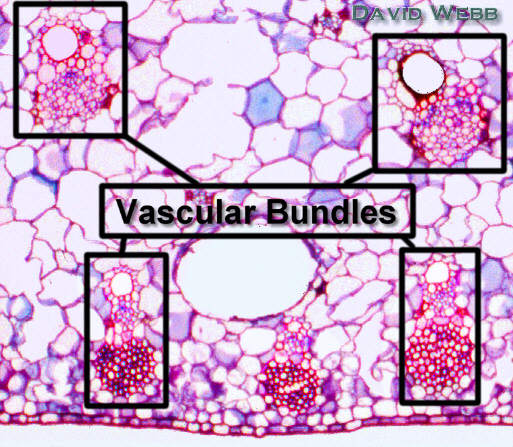
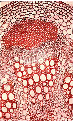
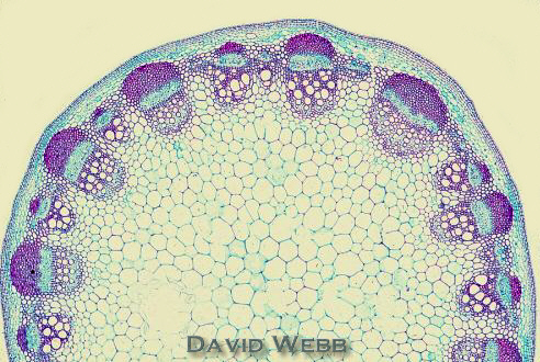
Observe
Dicot
stems usually have one ring of vascular tissue
in stems. The vascular cylinder is usually composed of individual vascular
bundles.
Study Helianthus (sunflower) stems in two stages of development.
The
epidermis is typical and stomata may be present.
The cortex is composed of parenchyma with abundant intercellular spaces.
Discrete vascular
bundles occur in the young stem.
Note the immature fiber bundles towards the outside of the phloem. These fibers
arise from the same part of the procambium as the primary phloem. The fibers
eventually develop lignified secondary walls.
T
Compare
Secondary vascular development is limited or absent in Helianthus. However, the vascular cambium may spread to the interfascicular areas. The fascicular and interfascicular cambia can unite and form a continuous ring which produces cylinders of secondary xylem and phloem. The primary xylem is displaced by the secondary xylem and is pushed into the pith.
The pith
is composed of parenchyma cells.
Study
hand sections
Stain
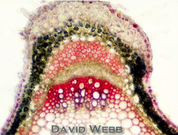
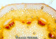 Widelia Stem stained with Phloroglucinol |
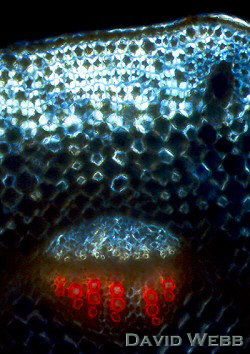 Widelia Stem stained with Phloroglucinol & viewed with crossed Polarizers |
Compare
Do the same for Coleus or another member of the Lamiaceae (Mint Family)
|
|
Cross section of Coleus stem stained with Phloroglucinol |
|
Study
|
|
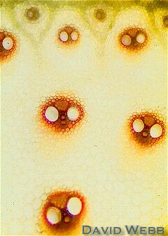
Sugar Cane Stem stained with Phloroglucinol |
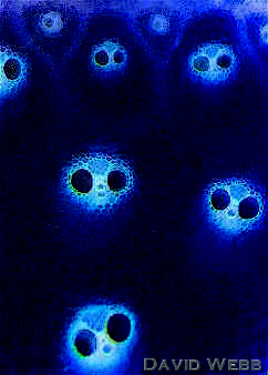
Sugar Cane Stem seen with Crossed Polarizers |
How does the arrangement of vascular bundles compare with dicot stems?
Are the bundles collateral, bicollateral, amphivasal or amphicribral?
Locate protoxylem and metaxylem? How does the cavity in the xylem develop?
Try to identify the sieve tubes and companion cells in the phloem. Locate the fibers associated with the vascular bundles.
|
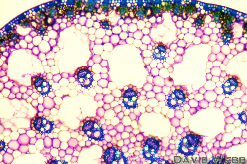 |
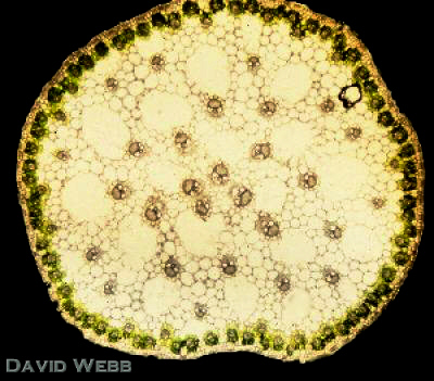
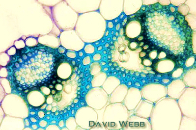
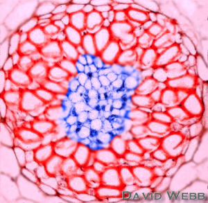
Cross Section of Makaloa stem: Note the widely distributed Vascular Bundles |
Makaloa stem stained with Toluidine Blue |
Examine cross sections
View unstained sections and sections stained with Toluidine Blue.
Note the widely distributed vascular bundles. This is similar to bamboo and sugar cane.
|
|
Air cavities are present in Makaloa which is a semi-aquatic plant. What term is used to describe tissue like this?
Makaloa used to make mats for the Hawaiian ali’i.
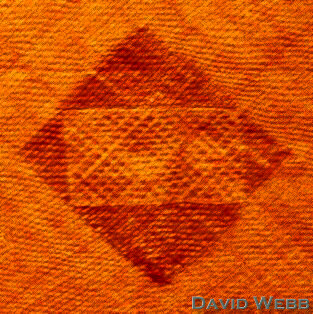
Piece of a Makaloa Mat displayed at the Bishop Museum: The symbol means hale
(house)
The whole stem was used to weave the mats.
The mats have a smooth & spongy
quality which makes them good for sleeping.
What anatomical features of makaloa account for the above qualities?
Examine the demo slide of an immature kalo (Taro) stem.
Note the abundance of parenchyma in this stem.
There is little starch in this slide because the stem was so young when it was harvested.
Locate the vascular bundles and note their distribution.
Is this plant a dicot or a monocot?
| Cross section of a young kalo stem: The area indicated by the box is shown at higher magnification below. |
|
|
|
|
|
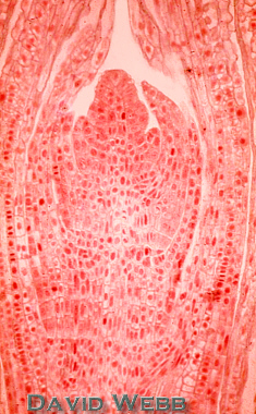
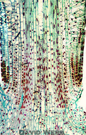
Many monocots have Intercalary Meristems. Intercalate means to insert between. In this case a meristematic layer occurs BETWEEN the Node and the Apical Meristem.
The Apical Meristem produces a small number of derivative cells towards the base. Those closest to the apical meristem enlarge and differentiate. Those more distal continue to divide and produce derivatives towards the apical meristem. This is the Intercalary Meristem.
These occur just above the nodes and function for a limited period and are thus said to be determinate. They are similar to the basal meristem which produces the strap like leaves of monocots. These are responsible for the re-growth of grass after it is mowed!
Examine
Locate the youngest nodes follow the cell files acropetally (towards the tip).
You should see that the least differentiated cells near the node and the most differentiated cells towards the apex.
Study the Illustrations
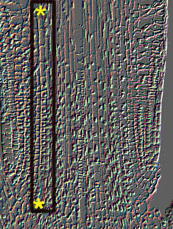
Follow the cell files indicated by the stars.
|
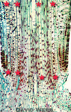 |
| Node-Internode of Equisetum: Follow the Cell Files from Bottom to Top (Acropetally) | |
|
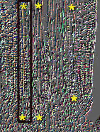 |
| Embossed Images of Equisetum Node-Internode | |
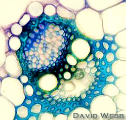
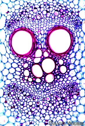
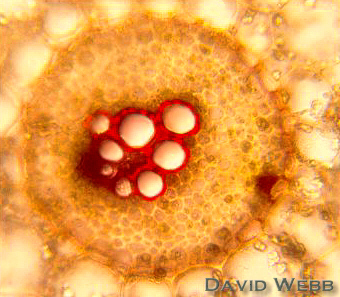
Vascular Bundle Types
Vascular bundles are classified according to the spatial relationships of their xylem and phloem.
Collateral bundles (Widelia, Helianthus, Coleus, Saccharum, and Cyperus) have Xylem on one side and Phloem on the other side. Bicollateral bundles (Cucurbita) have phloem on both sides of the xylem.
|
|
|
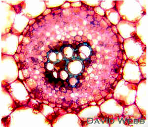
Concentric: There are two types of concentric bundles
amphicribral
amphivasal
|
 |
|
Amphicribral Bundle stained with Phloroglucinol |
Amphicribral Bundle stained with Toluidine Blue | Amphivasal Bundle with commercial staining |
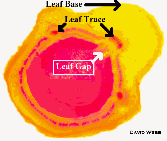
Nodal Anatomy
The study of nodes
reveals the association between the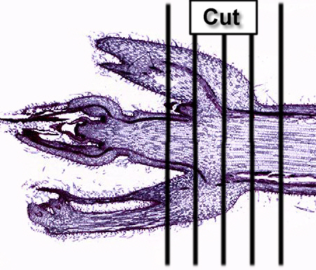 vascular
system of the stem and the vascular bundles of the leaves. The
part of a vascular bundle in the stem that diverges from the vascular cylinder and enters
the leaf is called leaf trace. In the location where the leaf trace is bent towards
the leaf, a relatively wide parenchymatous area appears in the vascular cylinder of the
stem. This area is the leaf gap. Different plants have different numbers of leaf
traces per leaf and different number of leaf gaps per node.
vascular
system of the stem and the vascular bundles of the leaves. The
part of a vascular bundle in the stem that diverges from the vascular cylinder and enters
the leaf is called leaf trace. In the location where the leaf trace is bent towards
the leaf, a relatively wide parenchymatous area appears in the vascular cylinder of the
stem. This area is the leaf gap. Different plants have different numbers of leaf
traces per leaf and different number of leaf gaps per node.
Make serial sections
Cut
These transverse sections must include some of the internode above and below the node as well as the node itself.
Arrange
Observe
Stain
Remember that the procambium forms a complete circle above the level of leaf initiation in some shoot apices.
The circle becomes interrupted as leaves are formed and develop.
These interruptions are leaf gaps.
The dissected vascular cylinders of dicots arise in this fashion.
Observe the demo slide of a Coleus shoot tip cut transversely in the region of leaf initiation.
Locate the ring of procambium and the leaf gaps!
 Cross Section through Coleus Shoot Tip |
Coleus has opposite leaves. Thanks a lot Drill Sargent!!!!!!!
![]()