Laticifers are some of the most peculiar elements of the plant body and are found in a restricted number of famous or infamous families and genera. Their infamy stems from the poisonous or addictive nature of their contents. However, the soothing qualitiies of the latex from the opium poppy led to the development of powerful pain killers like morphene which have made life and death more tolerable for seriously afflicted patients. Laticifers are long cells or vessel-like series of cells permeating various tissues of the plant. They contain variously colored, often milky juice called latex.
Galery of Familiar Plants which have Laticifers.
I have grouped these according to Families. I am not concerned that you learn these families or these plants by name. I want you to recognize plants that you see on campus and elsewhere in Hawaii. If you develop an interest in these plants you will want to learn about them for your own fulfillment.
Apocyanaceae
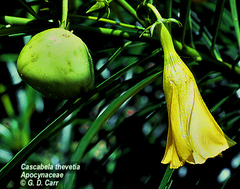 Cascabela thevetia |
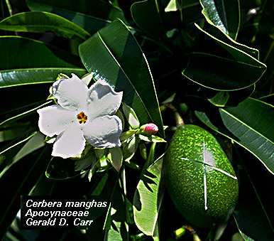 Cerbera manghas Note the latex oozing from the X cut into the fruit. |
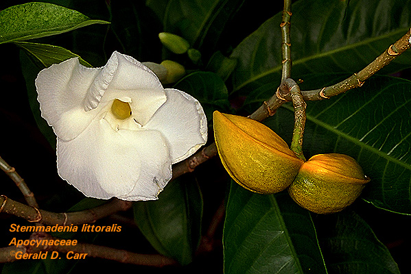 Stemmadenia littoralis |
|
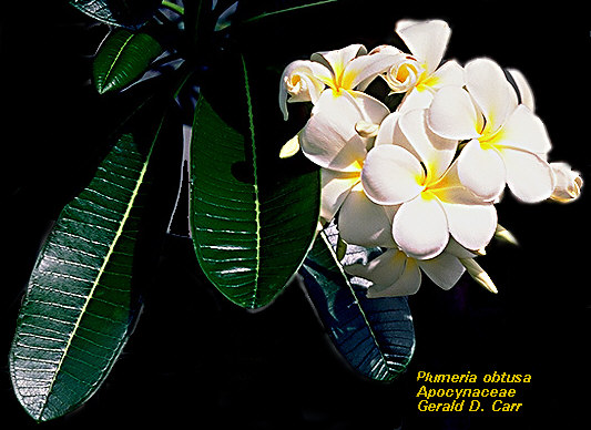 Plumeria obtusa |
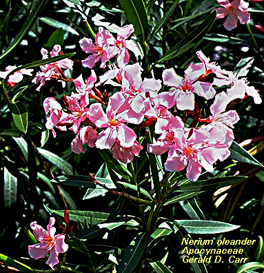 Nerium oleander |
||
Asclepiadaceae
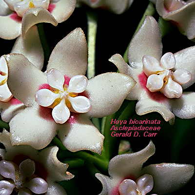 Hoya bicarinata |
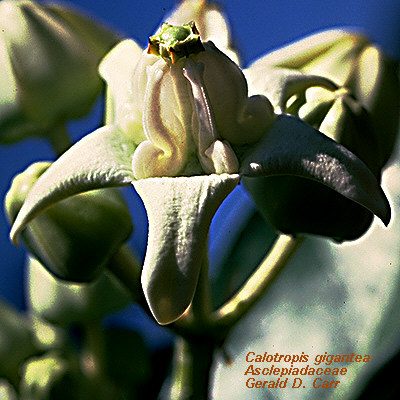 Calotropis gigantea |
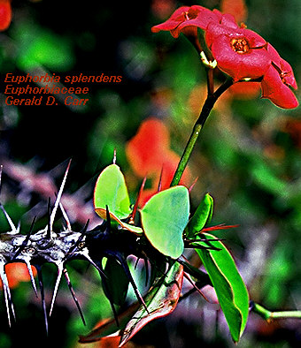
Euphorbiaceae
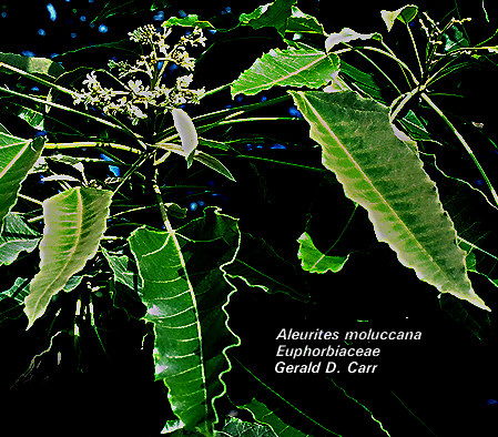 Aleurites moluccana |
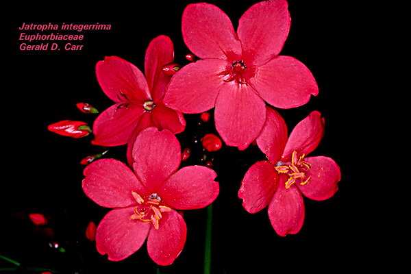 Jatropha integerrima |
|
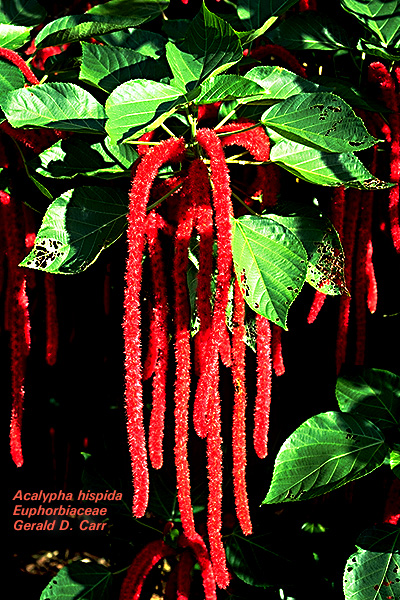 Acalypha hispida |
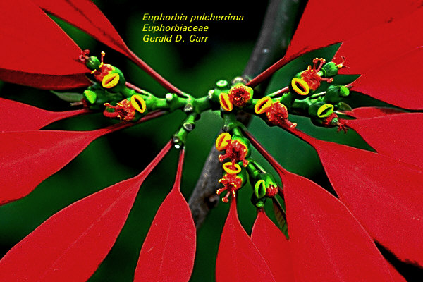 Euphorbia pulcherrima
|
|
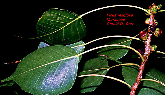
Moraceae
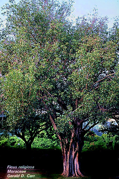 Ficus religiosa |
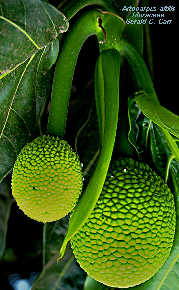 Artocrpus altis
|
Papaveraceae
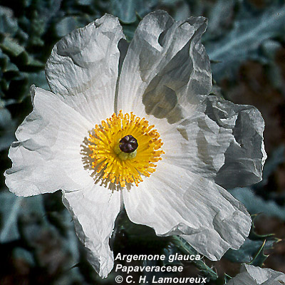 Argemone glauca |
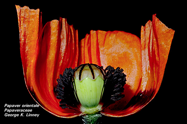 Papaver orientale |
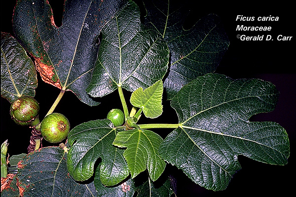
Examine the color of latices from various plants which may include Poppy (Papaver); Papaya (Carica papaya); Breadfruit (Artocarpus sp.); Euphorbia pulcherrima (white); Nerium oleander (colorless); Ficus sp. (milky white); Calophyllum inophyllum (yellow), (Casabella thevetia) or Plumeria.
Make Longitudinal sections of stems and petioles containing laticifers.
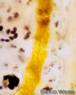
Stain these with IKI or Sudan
|
|
|
|
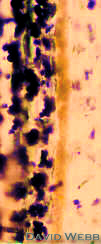
Laticifers stained with IKI |
Laticifers stained with Sudan |
||
Laticifers
in olona (Touchardia latifoliolata): 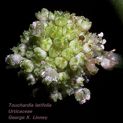
This plant produces incredibly strong "fibers" which were used by ancient Hawaiians to fashion their capes and helmets. Olona also produced the finest rope for sailing vessels and was unparalleled for its strength and durability until the introduction of synthetic polymers. The "fibers" are actually laticifers with incredibly thick walls.
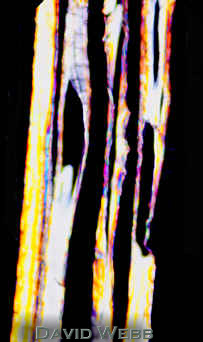
Make transverse and longitudinal sections of young stems.
View unstained and look for latex.
Examine them with your polarizers.
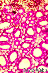
Stain
|
|
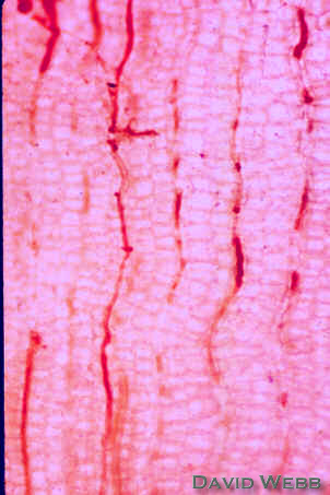
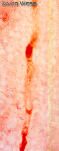
| Low Mag. View of Olona Stem seen with Polarized Light: Where are the thick-walled cells? | Unstained Laticifers at High Mag: Note the orange Latex in one laticifer, and the thick, translucent walls |
 |
 |
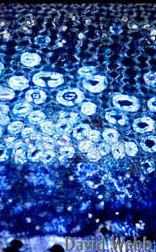
Longitudinal section of Olona stem viewed with crossed polarizers: What does the birefringence tell you about these cells? |
Olona Laticifers stained with Toluidine Blue |
NECTARIES
Floral Nectaries
These occur in a wide variety of locations on the flower. Some are easy to detect while others are not. We will consider only a few here. Dissect the following to find the nectariferous tissue, then examine available prepared slides.
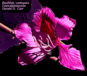
Bauhinia purpurea
The nectariferous tissue lines the opening of the tubular cavity subtending the petals, but only on the side opposite the large vascular bundle closest to the opening.
Locate the nectariferous tissue and vascular bundle first in fresh material, then look at the prepared slides.
The transverse sections of the receptacle show:
Only phloem occurs in the smallest bundles arranged in an area around the opening.
Phloem
ends in densely cytoplasmic cells.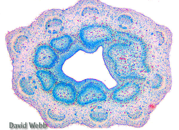
Larger, more vacuolate cells subtend the epidermis.
Epidermis and some other cells have yellow, granular contents.
Epidermis near the largest bundle has stomata but no cuticle.
The
nectariferous cells are the dense, small cells in the lower end
of the floral tube wall, just above the ovary. 
Carefully cut cross-sectional chunks off the tube, starting at the top, to see how much of the tube (of an upright flower) is filled with nectar.
A median
longitudinal cut should reveal the cells secreting the nectar.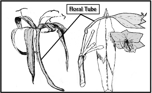
Acerola
Locate the floral nectaries with a dissecting scope.
Ring Nectaries
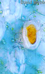
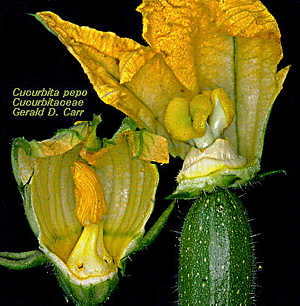
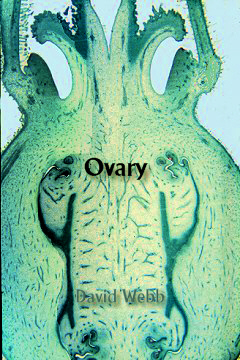
Many flowers have a "disk" or ring nectary around the base of the gynoecium, e.g., species of Asystasia, Alamanda, Convolvulus and Cucumis
Examine
the prepared slide of a median longitudinal section of Cucumis and find the nectary.
Can you deduce where the nectar is secreted?
Does the epidermis have a cuticle in this region?
Are both xylem and phloem present in the nectariferous tissue?
|
|
Locate similar nectaries on the corresponding living flowers.
 Allamanda sp. |
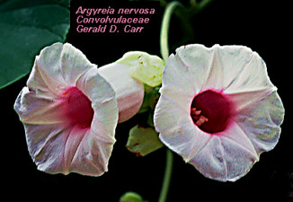 Argyreia nervosa (Convolvulaceae) |
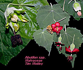
Abutilon
The
floral nectaries of various species from this genus and family (Malvaceae)
have been studied more than those of most plants. 
They are located on the upper surface of the base of the sepals, the secretory apparatus consists of closely packed hairs along with underlying nectariferous tissue.
To see nectar secretion in action, cut a thin transverse section of a sepal through this region and mount in immersion oil.
Look for small droplets of nectar at the tips of the hairs, and watch them grow.
Look also
at the prepared slide and locate the large
mucilage cells, druse cells & vascular bundles.
The vascular bundles are on the abaxial (lower) side, among, the
densely stained nectariferous cells.
Note that sieve tubes branch from the main vein and permeate the
nectariferous tissue.
|
|
Extrafloral Nectaries
These usually occur somewhere on leaves, but may be located on stems or bracts.
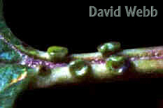
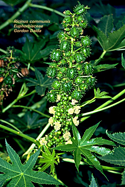 Ricinus communis
Find the two large nectaries on the petiole of a fresh leaf.
Observe a prepared slide that shows the densely stained nectariferous tissue.
Locate the tracheary elements and determine how close they come to the epidermis. Can you find sieve elements? Do they come closer to the epidermis? Note how the (secretory) epidermal cells are much denser than the two cell layers just underneath. Note also the thick cuticle covering the epidermal cells.
|
|
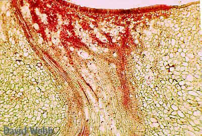
Extrafloral Nectaries of Ricinis communis |
|
Kuikui Leaf (near base of lamina),
 |
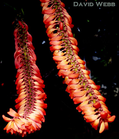
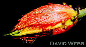
Norantea (floral bracts modified into large orange tubes)
|
|
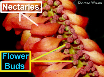
Norantea Inflorescence: The large Orange-Red structures are vase-like Nectaries & the Flower Buds are the small red spherical structures. |
|
Passiflora (floral bracts & petiole)
Impatiens (petiole)
Impatiens also has a floral nectary. Can you find
it?
kukui (Aleurites moluccana)
This plant plays an important role in Hawaiian culture. Ancient Hawaiians used virtually every part of this plant as an adhesive, for lamp oil, as waterproofing, as polish for storage vessels, in fire making, in lei making, in kappa making and in healing. It is still used to make fine leis. Many of its useful features are derived from the "oil" which can be obtained from its stems, leaves and fruits. Your challenge is to determine what type of secretory structure produces this oil!
You will examine hand sections to solve this puzzle.
Study unstained transverse and longitudinal sections from the petiole near its junction with the leaf blade.
Answer the following questions to solve the riddle.
Transverse Sections: Locate the red-brown areas.
In what regions or zones are they located?
What accounts for their color?
Are they one cell wide or many cells wide?
Are the secretory cells greatly enlarged compared to surrounding cells?
Is there evidence for a secretory epithelium?
Longitudinal Sections
Are the secretory cells rotund, truncated (short & rectangular) or highly elongated?
Do they form discontinuous secretory cavities or elongated, continuous channels?
Are they branched or un-branched?
Identify the Mystery Secretory Structure!!!!!!
![]()