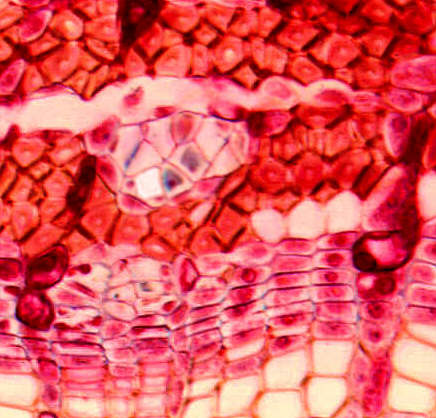
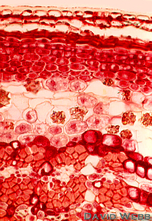
Secondary Phloem has the same origin
as secondary xylem, namely, the vascular cambium. Cells displaced towards the
outside of the vascular cambium differentiate as phloem. Secondary phloem can remain
active over several growth cycles. Secondary phloem, like secondary xylem, is a complex
tissue. It always has sieve elements which are analogous to tracheary elements.
However, parenchyma is also present, and sclerenchyma may also be visible.
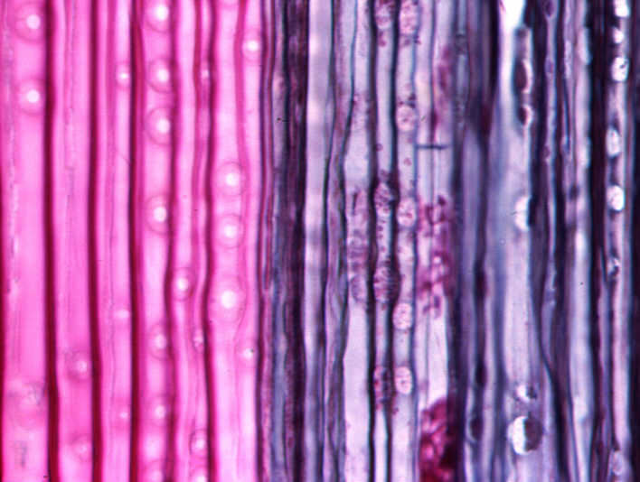
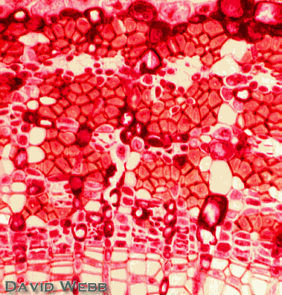
There are two kinds of sieve elements, Sieve Cells and Sieve Tube
Members.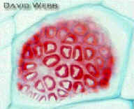
Sieve Tube Members are highly specialized for translocation. They are moderately elongate and have horizontal or oblique end walls with sieve plates. Sieve plates contain large sieve pores. The pores are lined with callose which regulates the diameter of the pores. Callose plugs the pores when the cells are ruptured.. The pores can also be closed slowly in response to major changes in the environment, like the onset of winter in the Temperate Zone. Pores closed in this manner may open when growing conditions become favorable. Sieve tubes are formed by the vertical union of several sieve tube members.
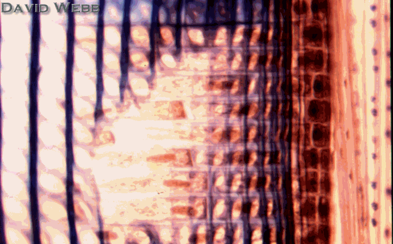
![]() Sieve Cells
are also specialized for translocation but they lack sieve plates and have a
narrower diameter. However, sieve pores are more numerous where sieve cells overlap. Callose
is also associated with the sieve pores. Sieve cells are more highly elongated
than sieve tube members.
Sieve Cells
are also specialized for translocation but they lack sieve plates and have a
narrower diameter. However, sieve pores are more numerous where sieve cells overlap. Callose
is also associated with the sieve pores. Sieve cells are more highly elongated
than sieve tube members.
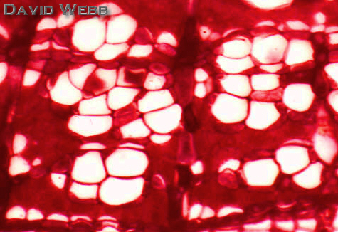
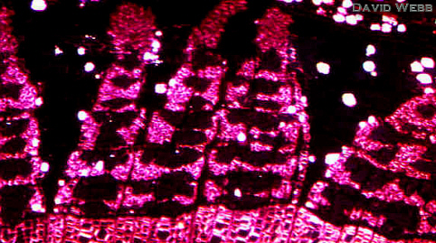
The cell walls of most sieve elements are similar to those of parenchyma cells, and it is often difficult to distinguish between these in cross sections unless a sieve plate or sieve pores can be seen. Callose is preferentially stained by aniline blue. Consequently, sieve elements may be located by their reaction to the stain. However, other cells also react positively with this stain. Callose stained with aniline blue fluoresces under near ultra violet and violet light. This provides a more certain method to locate sieve elements. Callose is also present in their cytoplasm and its fluorescence aids in the location of sieve elements.
Sieve tube members have companion cells, and sieve cells have albuminous cells associated with them. These are also hard to distinguish from phloem parenchyma, especially in cross sections. Starch is usually absent from albuminous cells and this can provide a way to distinguish them.
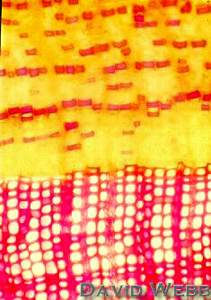
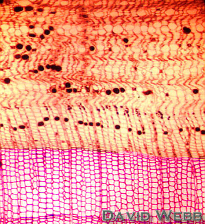
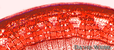
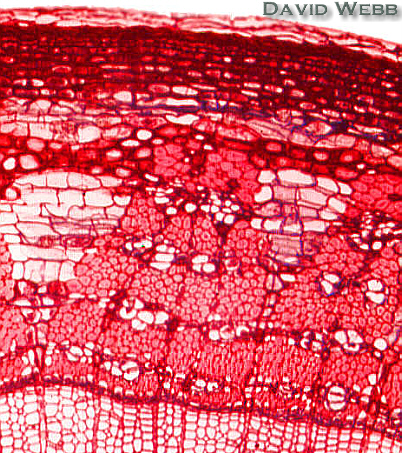
Rays are present in secondary phloem and are usually continuous with rays in the secondary xylem. Some species develop dilated rays in their secondary phloem. Tilia americana and Hibiscus tiliaceus (hau) are two prime examples for the presence of dilated rays. Dilated rays are principally composed by parenchyma cells which result from localized anticlinal divisions.
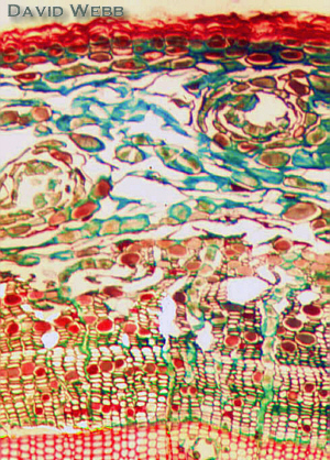
Secondary Phloem of Gymnosperms
You should recall that the secondary xylem of gymnosperms like Norfolk Island Pine is simple compared to angiosperms like (Hibiscus tiliaceus). The same is true for secondary phloem.