 |
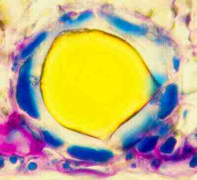 |
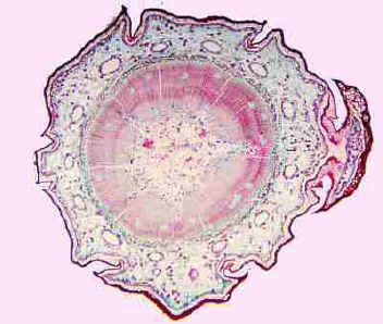
1] Identify this Entire Structure Secretory Cavity or Canal 2] Is it Lysigenous or Shizogenesis Shizogenousor Lysigenous 3] How do you justify your choice for #2???? Epithelium present or Absent |
|
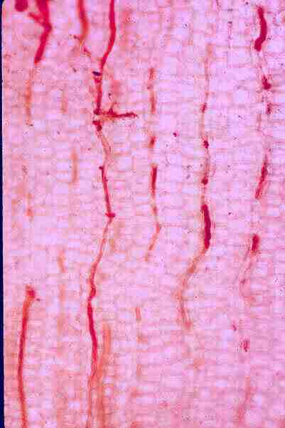 |
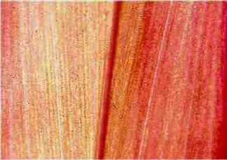
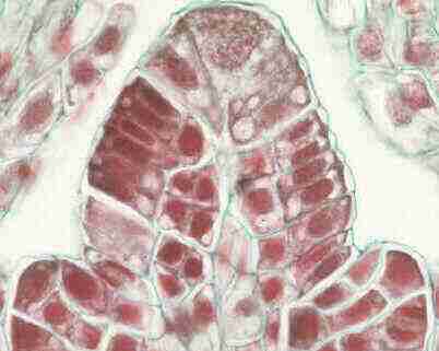
4] Identify these long structures that have stained Red Laticifer 5] What Stain was used? Sudan |
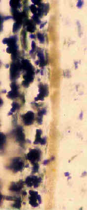 |
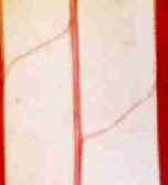
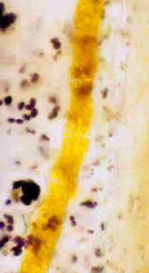 6] What Stain was Used? IKI 7] Identify the cell which has stained Yellow Laticifer |
 |
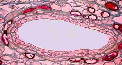 8] Identify the Cells which line this area Epithelium 9] Is it Lysigenous or Shizogenous? |
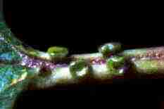 |
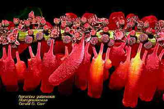
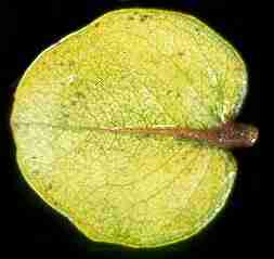
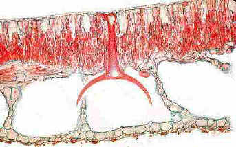
10] What General Term is used to describe structure like this? Nectary 11] What 2 terms would be used to Specifically classify this kind of structure? Extrafloral, Structural |
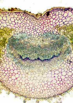
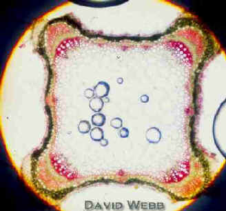
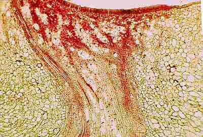 This is a micro section of the structure above! 15] Identify this tissue Phloem or Vascular 16 [Bonus] What Global term is used to describe both of these Tissues in structures like this? Nectariferous
|
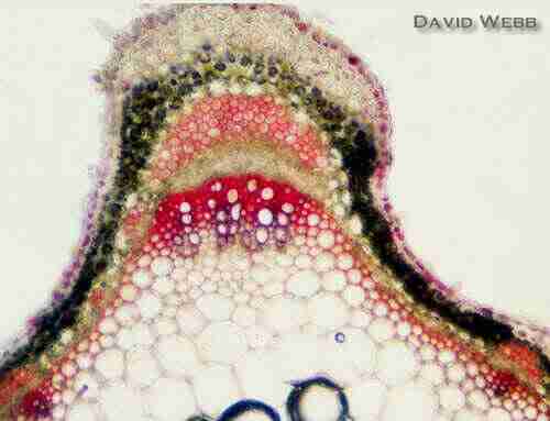
12] Identify the tissue on the surface Epidermis 13] Do these cells differ from their counterparts in this area? YES! 14] What does this suggest about their specific function in this area? Secreting Nectar
|
 |
17] Identify these
Structures 18] Identify these Structures |
 |
 |
 |
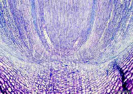
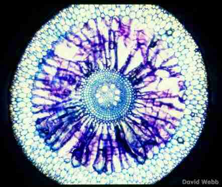
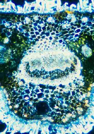
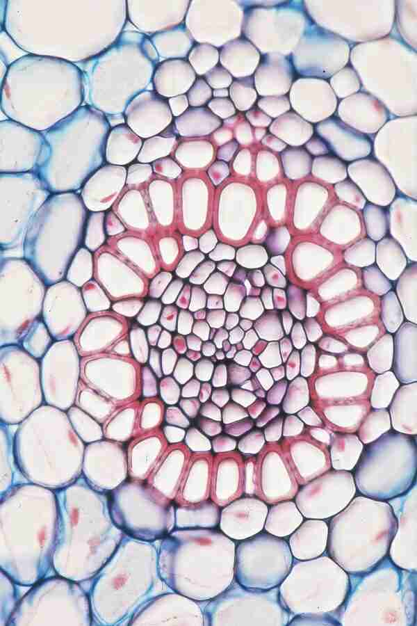
19] Identify the Organ 20] What General Term is used to describe this entire area? 21] What specific Term is used to describe this Tissue. |
 |
22] What Stain was used? Toluidine Blue 23] Identify this Tissue Phloem 24] Identify this Tissue Xylem |
25] Identify this Red
Band 26] Identify the Unicellular Layer in which it is typically found. |
|
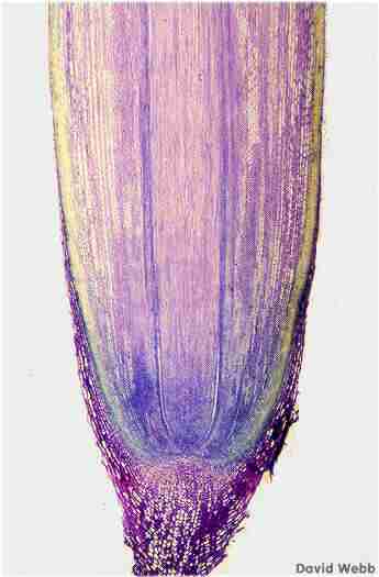 30] What general type of tissue develops in this area? Vascular or Pith 31] What general type of tissue develops in this area? Ground |
 27] Identify this Structure Root Cap
28 [Bonus] Specifically identify the Meristem that produces this structure. Calyptrogen 29] Is this Open or Closed? |
|
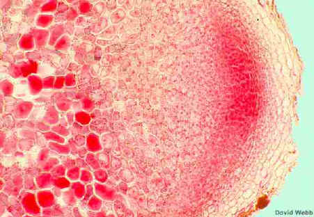
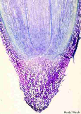
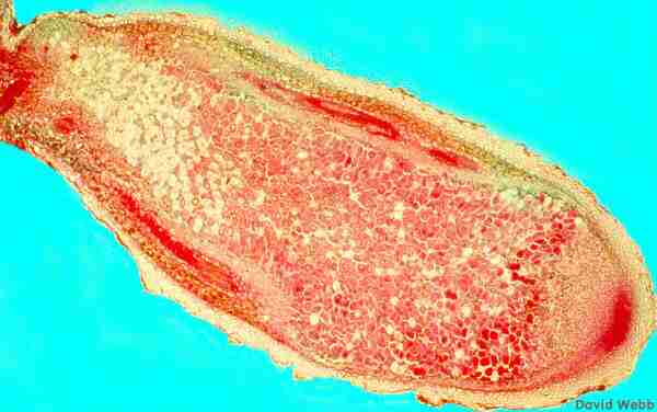
32] What general Term is used to describe a structure like this? Root Nodule 33] What General Term is used to describe an area like this? Apical Meristem 34] Why are these cells heavily stained? Contain Bacteria 35] What is the function of this structure Fix Nitrogen 36] BONUS] What term is used to describe the longevity of structures like this Indeterminate |
 |
|
 |
 |
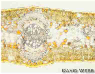
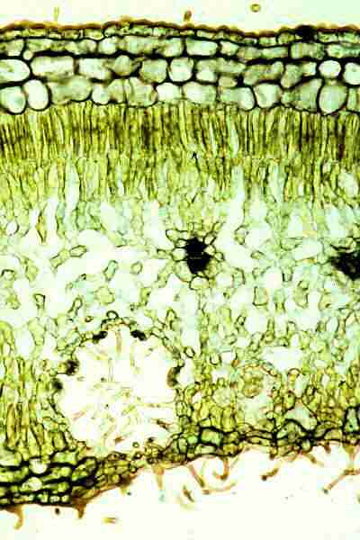
37] What part of the leaf is represented? Midrib 38] Identify this Tissue Xylem 39] Identify this Tissue Phloem |
|
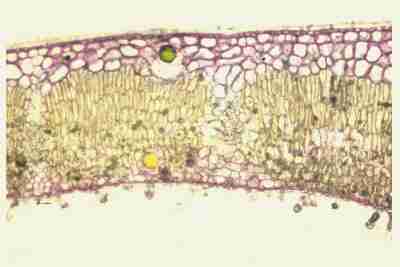 |
40] Specifically Identify this region. Hypodermis 41] Identify this Area Palisade |
 42] Identify this Area 43] Identify these Cells 44] Identify these Cells 45] Identify this Area Stomatal Crypt |
 |
|
46] Identify this Tissue Mesophyll 47] What is its Function Photosynthesis 48] Identify this Unicellular Layer Endodermis 49] Specifically identify the Tissue in this region Transfusion 50] What specialized cells are found in this tissue Tracheids |
 |
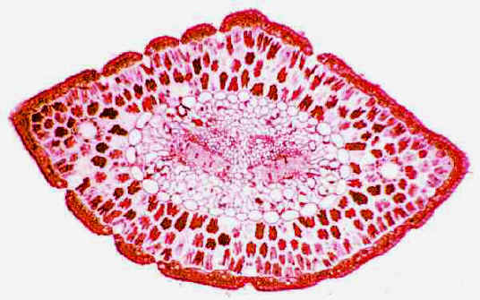
51] Is this Bifacial or Unifacial? 52] [BONUS]What part of the Juvenile Leaf is represented here? Rachis or Petiole or Midrib |
  |
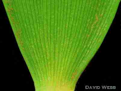
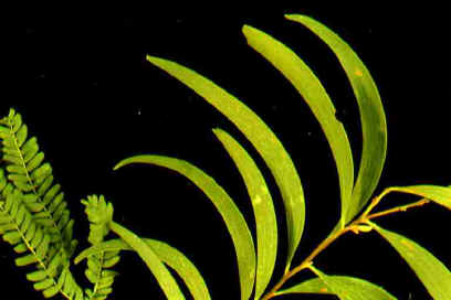
53] What type of Venation is Illustrated Striate or Parallel
54] BONUS] Specifically Identify this Structure Commisural Bundle |
  |
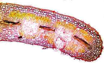
55] What stain was used Phloroglucinol 56] Identify this Tissue Fibers (Phloem) 57] Identify this Tissue Primary Xylem 58] Identify the Tissue in this region
|
|
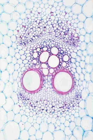
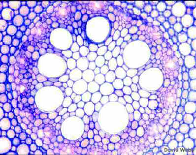
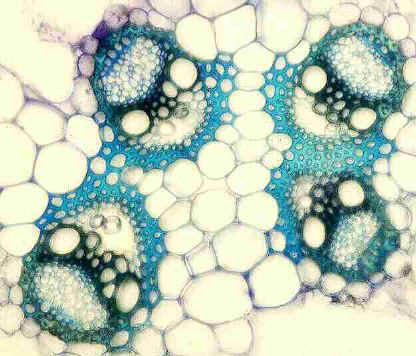
59] Is this a Monocot or a Dicot 60] What Stain was used? Toluidine Blue 61] What kind of Vascular Bundle is this Collateral (Monocot) 62] Identify these circular Structures Vascular Bundles 63] Identify this Tissue Fibers
|
 |
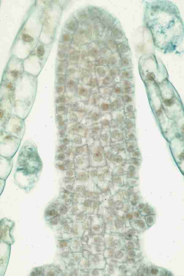
64] What Type of Apical Meristem Organization is illustrated Apical Cell |
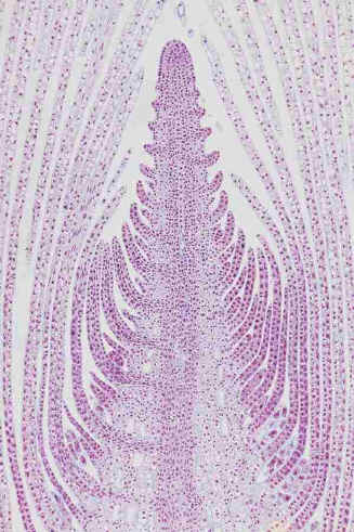 65] Identify these structures Leaf primordia |
67] Identify its counterpart which would be found in this
region. |
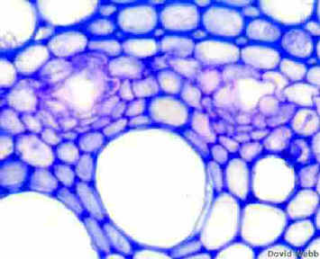
 68] Specifically Identify this Type of Vascular Bundle Amphivassal |
|
| 70] BONUS] Identify the
Type of Venation Dichotomous
|
|
 |
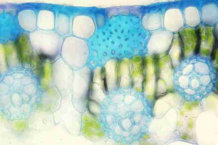
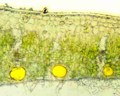
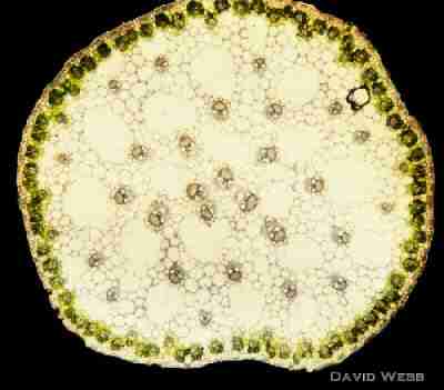
71] Identify this Red-stained Tissue Chlorenchyma or Photosynthetic Parenchyma or Palisade 72] Identify the structures which lie above these cavities Stomata 73] Identify this Tissue Aerenchyma 74] Is this commonly found in plants which grow in Hydric, Mesic or Xeric Environments? |
![]()