![]() Fluorescence Microscope Images
Fluorescence Microscope Images ![]()

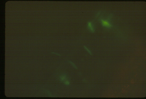
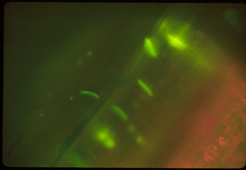
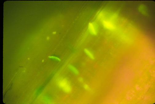
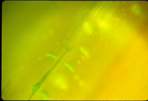
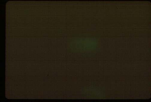
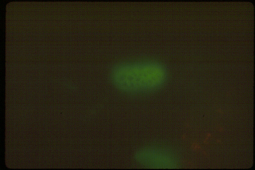
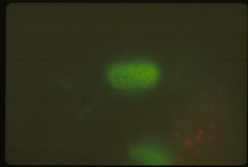
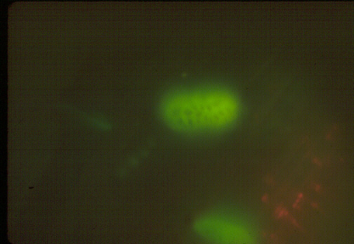
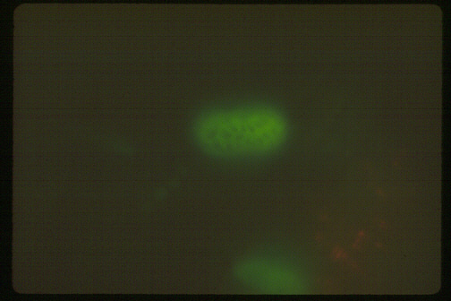
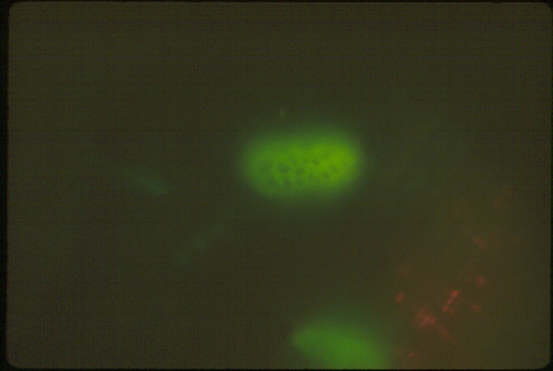
Cucumber Phloem stained with Aniline Blue & Viewed with Violet Light Fluorescence.
| Longitudinal
sections of Cucumis stems were stained with Aniline Blue and
photographed with a fluorescence microscope after excitation with Violet
Light. Callose in the Phloem is fluorescent and appears bright under these conditions. It has a yellow/green color. This is partly due to the blue color of the dye. The Sieve Plates are especially fluorescent because they contain a lot of associated Callose and Callose accumulates there upon injury. Lateral Sieve Pores are also fluorescent. Xylem is autofluorescent and appears similar to the Phloem. Xylem can be identified because of its characteristic secondary walls. Plastids appear a red dots due to the fluorescence of Chlorophyll
There have been no modifications of the raw scanned images!!!
The times below each photo indicate the time that the shutter was OPEN! |
|
| 20X Magnification | |
|
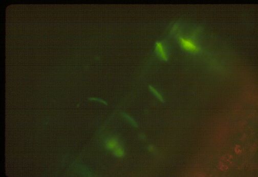 2 Seconds |
|
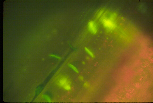 8 Seconds |
|
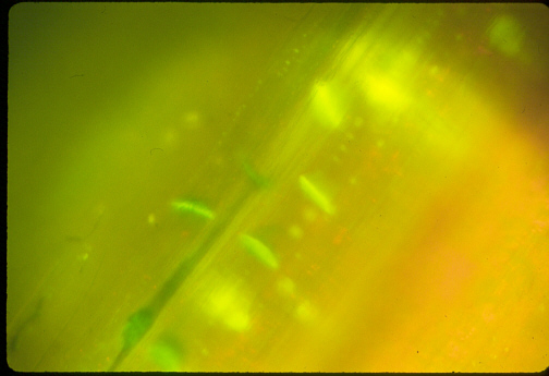 30 Seconds |
|
|
40X Magnification of a Sieve Plate |
|
|
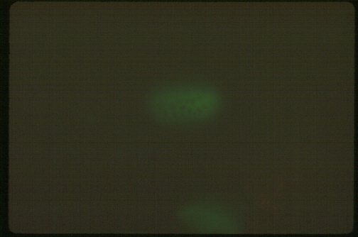 2 Seconds |
|
 8 Seconds |
|
 30 Seconds |
|
 120 Seconds |