
The Pterophyta is a familiar
plant taxon,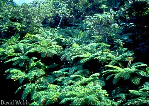 especially in Hawaii. They have a prominent position in our terrestrial ecosystems both as
ground cover species and canopy components in certain environments. They are an order of
magnitude larger than the plants we have reviewed so far. However, even Tree Ferns are
relatively small plants compared to woody Angiosperms and Gymnosperms.
especially in Hawaii. They have a prominent position in our terrestrial ecosystems both as
ground cover species and canopy components in certain environments. They are an order of
magnitude larger than the plants we have reviewed so far. However, even Tree Ferns are
relatively small plants compared to woody Angiosperms and Gymnosperms.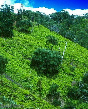
We will concentrate on the Filicales which contains most extant species. They are most abundant in the tropics but some species can be found in the arctic alpine zone.
Ferns typically have subterranean stems called Rhizomes. Some species have Stolons. Most species are terrestrial but some are epiphytic and a few are aquatic.Some species produce significant aerial stems and resemble small trees. Hence, their designation as "Tree Ferns".
Roots are adventitous and arise
from 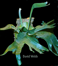 the stem. The stems of
tree ferns have a thick coat formed by adventitous roots which are initiated near the apex
of the stem and grow down the outside of the stem. This has some obvious disadvantages.
The roots must travel a long distance through the atmosphere before reaching the soil.
Consequently, water loss can readily occur over this distance. They are also subjected to
any biotic or abiotic events that occur over this expanse.
the stem. The stems of
tree ferns have a thick coat formed by adventitous roots which are initiated near the apex
of the stem and grow down the outside of the stem. This has some obvious disadvantages.
The roots must travel a long distance through the atmosphere before reaching the soil.
Consequently, water loss can readily occur over this distance. They are also subjected to
any biotic or abiotic events that occur over this expanse.
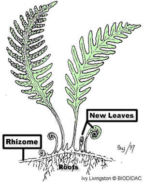 Diagram of a "typical" fern Diagram of a "typical" fern |
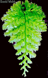
 Laua'e
(Male Scented Fern) |
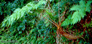 Hawaiian Tree Fern (Cibotium) along the Manoa Cliffs Trail. 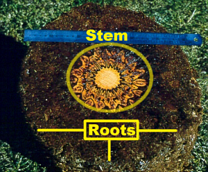 Cross section of a tree fern (Dicksonia) showing the thick mantle of roots that ensheath the stem. |
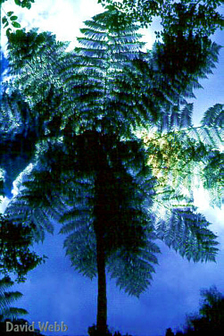 Tree fern from Puerto Rico. |
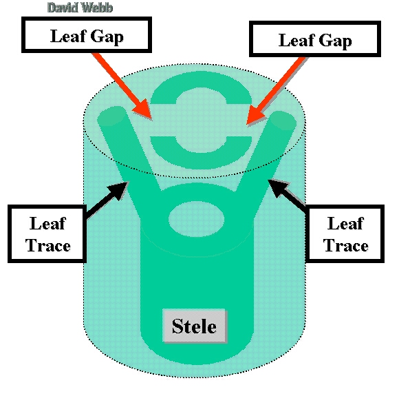
The most significant vegetative 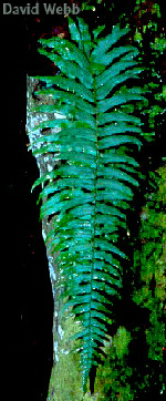 adaptation seen in
these plants is the Megaphyll (Big Leaf). However,
size is not the distinguishing trait for Megaphylls. A Megaphyll has
more than one vein in its blade and the Leaf Trace is associated with a Leaf Gap in the
Stem.
adaptation seen in
these plants is the Megaphyll (Big Leaf). However,
size is not the distinguishing trait for Megaphylls. A Megaphyll has
more than one vein in its blade and the Leaf Trace is associated with a Leaf Gap in the
Stem.
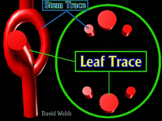
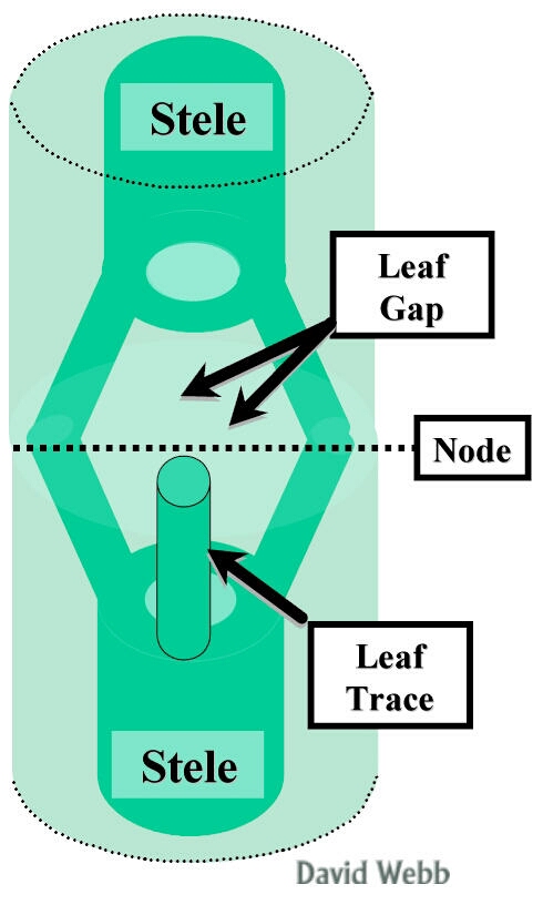
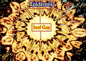
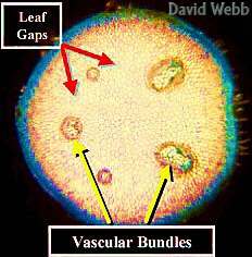
A Leaf Gap occurs when part of the vascular tissue in the stem is diverted towards the leaf. This is called a Leaf Trace. The integrity of the stele is restored above the Leaf Gap. When separate vascular bundles constitute the Stele, they may fork to produce Stem Traces which unite above the Leaf Gap to reform the Vascular Bundle. Parenchyma cells replace the Leaf Trace above the level of its divergence. Consequently, if you make a series of cross sections of a stem, it will appear that a gap occurs in the Stele near the level where the Leaf Trace diverged. The Diagrams below attempt to depict ways in which this could occur.
  |
 |
I will try to show you how leaf gaps occur in the table below.
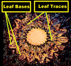
This fern (Dicksonia) has a Siphonostele which has a solid cylinder of Vascular Tissue. Part of the Stele diverges into the leaf bases which are clustered around the circumference of the stem. The leaf traces are V-shaped and the entire profile of the Stele and the Leaf Traces resembles a sun and rays emanating from it. The Leaf Gaps are the light areas that intervene the circular outline of the stele. |
 |
 |
 |
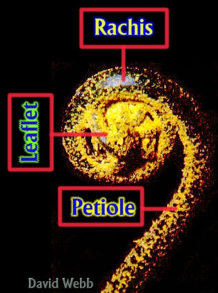
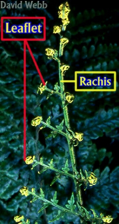
The predominate organizational pattern for fern leaves is Pinnate.This means that a structure has one central axis which produces lateral structures that are opposite each other along the central axis. A bird feather is a good example of a structure that has pinnate organization. Pinnate leaves can be Simple, Compound and Bicompound. The central axis of compound leaves is called the Rachis at the level of Leaflet production. The leafless portion that joins the stem is the Petiole.
The presence of Megaphylls and their associated Leaf Gaps in the Stele result in the occurrence of complex stelar organization in some ferns. Leafy stems have dissected steles while Rhizomes (produce few or no leaves) have relatively undissected steles.
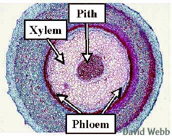
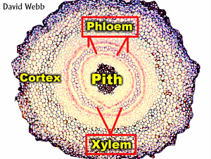
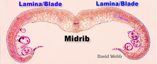
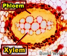
A Siphonostele has a Hollow Cylinder of Vascular Tissue which encloses a central Pith. The Cortex is the Ground Tissue between the Vascular Cylinder and the Epidermis.
 |
 |
| Simple Siphonostele with no Leaf Gaps. | Dicksonia Stem with a Siphonostele: A Leaf Gap is visible at 6:00. |
 |
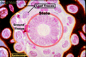 |
| Fern Stele that contains separate vascular bundles. This is a Dictyostele (Divided Stele) | Osmunda stem with a Siphonostele. The vascular bundles are leaf traces. The dark area outside of the Epidermis is composed of hairs. |
Leaf Venation (vein pattern) can be Dichotomous or Reticulate.
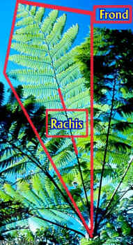 A Fern leaf is often called a Frond. A Rachis exists with Compound Leaves. It is an extension of the Petiole to which Leaflets are attached. In this case the Leaflets are also compound. Consequently, the leaf is twice compound (Bicompound.). |
||
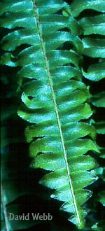
 Pinnate organization gone wild! |
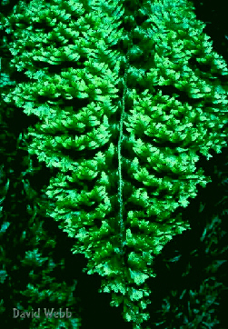
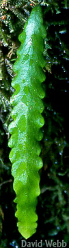
 A local Filmy Fern that has Pinnate organization. It is easy to see the complex venation in this leaf. |
|
 Pinnate Compound Leaf |
 Pinnate Simple Leaf |
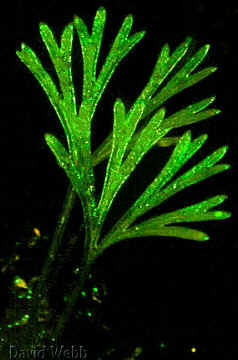 Some fern leaves have a Dichotomous organization. |
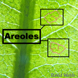 This fern leaf has Reticulate Venation. Two Areoles have been outlined in red. |
Close
inspection of the leaf below shows that it has Dichotomous Venation.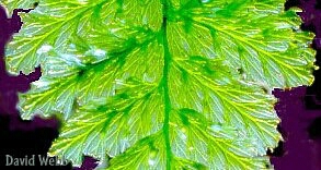 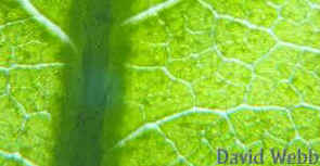 The image above shows Reticulate Venation. |
|
| Dichotomous Venation is
characterized by the repeated bifurcation (split in two) at the tip of each vein. This is
repeated over and over. Reticulate Venation is characterized by a more intricate pattern of branches which become progressively smaller and smaller until the surround a small area called an Areole. The ultimate branch vein terminates within the Areole. This is a Derived "Advanced" trait which is associated with flowering plants. Reticulate Venation is more specialized and is more efficient than Dichotomous Venation. |
||
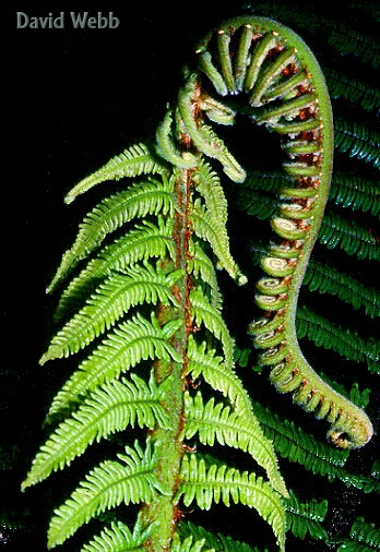
Fern Leaves have
a unique manner of development. The immature leaves are tightly coiled into a structure
called a "fiddlehead". During maturation, the leaf uncoils from the bottom to
its top (acropetal). This is called Circinate Vernation
(Circ = Circle; Verna = Spring).
 Tightly coiled Immature Leaf |
 Leaf tip from an uncoiled leaf of the same species. |
 Tightly coiled fiddlehead |
 Nearly mature frond with some coiled leaflets |
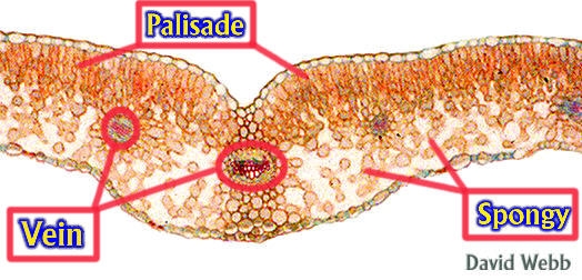
Leaf Anatomy can be simple or can be as complex as that seen with flowering plants. The take home message is that fern leaves are highly sophisticated in their design and represent a major advance over the Microphyll!
 |
 |
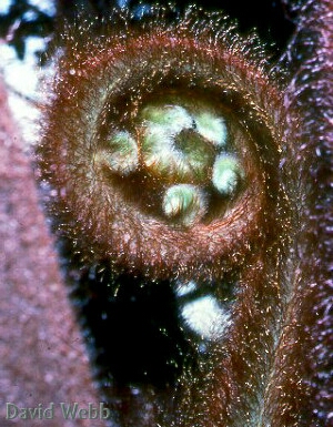
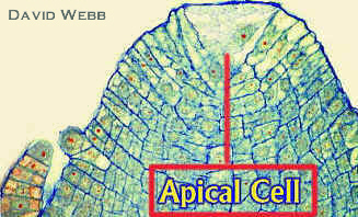
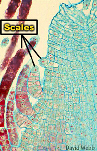
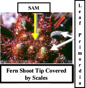
| Fern Apical Meristems tend to have Apical Cells. The small structures along the flank of the stem apex are Scales. These protect the immature leaves and the Shoot Apical Meristem. | |
 |
 |
 Fern Shoot Apex: The protruding structures are Leaf Primordia. Observe the labeled image to the right. |
 This fern grows in northern Massachusetts and these scales represent a significant adaptation in this climate. |
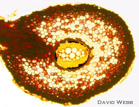
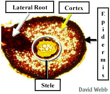
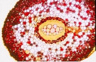
Fern Roots have the same basic anatomy as other roots. They do exhibit Lateral Branching which is a major advance over Dichotomous Branching.
| Cross section of Pteris Root with a Lateral Root | |
 |
 |
 |
 |
![]()