PALAEOBOTANICAL RESEARCH   GROUP
 
UNIVERSITY  MÜNSTER

THE   RHYNIE  CHERT  AND  ITS  FLORA

 
III. RHYNIA  AND  AGLAOPHYTON
 
Rhynia gwynne-vaughanii and Aglaophyton major which are traditionally classified within the Rhyniophytes look very similar, but differ clearly in their size and in the wall structure of the water-conducting cells.  The axes of Rhynia are up to 3 mm, whereas those of Aglaophyton can be up to 6 mm in diameter. Both species have a central stele consisting of water conducting cells surrounded by a tissue that conducted assimilation products. Most of the axis consists of cortex, which is differentiated into an inner cortex and an outer cortex where photosynthesis took place.  The cortex is surrounded by a hypodermis and an epidermis, the latter being covered by the cuticle.  New studies have shown that the conducting cells of Aglaophyton major are strongly reminescent of those of certain mosses; some authors therefore do not regard Aglaophyton as a real vascular plant. However, it should be kept in mind that in all other respects both forms are very similar. 
Cross section through an axis of Rhynie gwynne-vaughanii
 
Aglaophyton is the best known of all Rhynie Chert plants.  The plant was up to 15 cm high and consisted of naked creeping axes which were occasionally bent upwards and bifurcated.  Longer axes were bent down again, resulting in typical U-shaped morphology of the axes.  Especially in the lower parts these axes bore many lateral axes, which can be regarded as vegetative daughter plants.  The sinuous axes were lying loosely on the substrate surface and functioned as rhizomes.  Where the axes touched the substrate so-called rhizoids were formed.  These unicellular hair-like protrusions of the epidermal cells served for the intake of water and nutrients.  The entire plant was lying on the substrate. The stomata through which gas exchange took place, consisted of two kidney-shaped guard cells.  
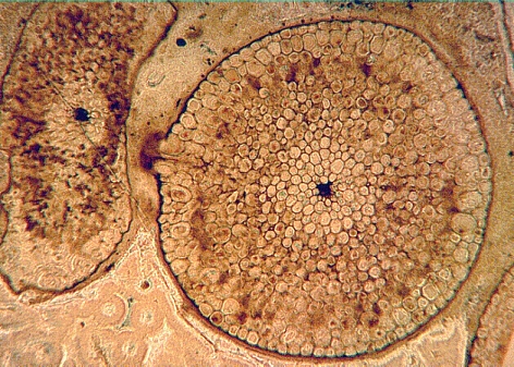
Photosynthesis  took place in the axes, like in other leafless plants.  Although no chlorophyll has been found, the special shape of the cells shows the location of the photosynthetic tissue.  The cells of the outer cortex are pallisade-like and directed upwards like in modern plants with upright standing photosynthetic axes or leaves, e.g. Juncus
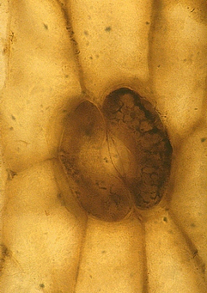
Aglaophyton had terminally attached elongate sporangia which opended with a spiral slit; the spores show a clear trilete mark.  
 
A stoma of Aglaophaton major with the two kidney-shaped guard cells


© Forschungsstelle für Paläobotanik, Westfälische Wilhelms-Universität Münster 
April 2000