
Botany online 1996-2004. No further update, only historical document of botanical science!
1. and 2. Electron microscopic pictures of dictyosomes from cultivated Acer pseudoplatanus cells. A to C Dictyosomes cut at different planes (preparations are 70 nm thick). 2. Fixing of the outlines. The scanned picture (see above) is digitalized and viewed on a screen. The outlines of interest have now to be fed into the computer (white lines in the picture). Both the original picture and the overlapping scheme are stored. Every electron microscopic picture has to be processed in this way.
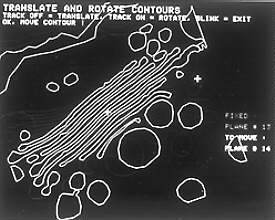
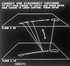
3. Depiction of the outlines (only these are worked on in the following). Now the outlines of the single layers are arranged with respect to each other by turning and shifting. These corrections have to be done because the co-ordinates of the series of sections are not co-ordinated. 4. Evaluation of the three-dimensional structure by making connections between the outlines of neighbouring layers.

5. A comparative model made from synthetic material. The outlines are copied to plates of synthetic material and resulting shapes were cut out. The single plates were used for the reconstruction of the three-dimensional structure of the dictyosome. In the pictures to the left and the right parts of the endoplasmic reticulum are depicted (striped). 6. The same set of data as in 5 was used. The reconstruction was done with a computer using steps 2 to 4 as a basis (W. MENHARD, J. LOCKHAUSEN, W. J. DALLAS, U. KRISTEN, 1986).