
Botany online 1996-2004. No further update, only historical document of botanical science!
In 1924 L. de BROGLIE discovered the wave-character of electron rays thus giving the prerequisite for the construction of the electron microscope. The prototype was built by M. KNOLL and E. RUSKA (Technische Universität Berlin, 1932). One of the first biological objects depicted was the tobacco mosaic virus (TMV). The first picture of a cell was published in 1945 by K. R. PORTER, A. CLAUDE and E. F. FULLAM (Rockefeller Institute, New York).
 The
conventional electron microscopy is nowadays called TEM (transmission
electron microscopy). We will therefore start with its construction.
The ray of electrons is produced by a pin-shaped cathode heated up by
current. The electrons are vacuumed up by a high voltage at the
anode. The acceleration voltage is between 50 and 150 kV. The higher
it is, the shorter are the electron waves and the higher is the power
of resolution. But this factor is hardly ever limiting. The power of
resolution of electron microscopy is usually restrained by the
quality of the lens-systems and especially by the technique with
which the preparation has been achieved. Modern gadgets have powers
of resolution that range from 0,2 - 0,3 nm. The useful resolution is
therefore around 300,000 x.
The
conventional electron microscopy is nowadays called TEM (transmission
electron microscopy). We will therefore start with its construction.
The ray of electrons is produced by a pin-shaped cathode heated up by
current. The electrons are vacuumed up by a high voltage at the
anode. The acceleration voltage is between 50 and 150 kV. The higher
it is, the shorter are the electron waves and the higher is the power
of resolution. But this factor is hardly ever limiting. The power of
resolution of electron microscopy is usually restrained by the
quality of the lens-systems and especially by the technique with
which the preparation has been achieved. Modern gadgets have powers
of resolution that range from 0,2 - 0,3 nm. The useful resolution is
therefore around 300,000 x.
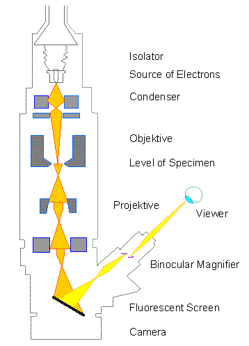
The accelerated ray of electrons passes a drill-hole at the bottom of the anode. Its following way is analogous to that of a ray of light in a light microscope. The lens-systems consist of electronic coils generating an electromagnetic field. The ray is first focused by a condenser. It then passes through the object, where it is partially deflected. The degree of deflection depends on the electron density of the object. The greater the mass of the atoms, the greater is the degree of deflection. Biological objects have only weak contrasts since they consist mainly of atoms with low atomic numbers (C, H, N, O). Consequently it is necessary to treat the preparations with special contrast enhancing chemicals (heavy metals) to get at least some contrast. Additionally they are not to be thicker than 100 nm, because the temperature is raising due to electron absorption. This again can lead to destruction of the preparation. It is generally impossible to examine living objects.
After passing the object the scattered electrons are collected by an objective. Thereby an image is formed, that is subsequently enlarged by an additional lens-system (called projective with electron microscopes). The thus formed image is made visible on a fluorescent screen or it is documented on photographic material. Photos taken with electron microscopes are always black and white. The degree of darkness corresponds to the electron density (= differences in atom masses) of the candled preparation.
 The
path of the electron beam within the scanning electron microscope
differs from that of the TEM. The technology used is based on
television techniques. The method is suitable for the depiction of
preparations with conductive surfaces. Biological objects have thus
to be made conductive by coating with a thin layer of heavy metal
(usually gold is taken). The power of resolution is normally smaller
than in transmission electron microscopes, but the depth of focus is
several orders of magnitude greater. Scanning electron microscopy is
therefore also well-suited for very low magnifications. Numerous
examples will be given in the following.
The
path of the electron beam within the scanning electron microscope
differs from that of the TEM. The technology used is based on
television techniques. The method is suitable for the depiction of
preparations with conductive surfaces. Biological objects have thus
to be made conductive by coating with a thin layer of heavy metal
(usually gold is taken). The power of resolution is normally smaller
than in transmission electron microscopes, but the depth of focus is
several orders of magnitude greater. Scanning electron microscopy is
therefore also well-suited for very low magnifications. Numerous
examples will be given in the following.
The surface of the object is scanned with the electron beam point
by point whereby secondary electrons are set free. The intensity of
this secondary radiation is dependent on the angle of inclination of
the object's surface. The secondary electrons are collected by a
detector that sits at an angle at the side above the object. The
signal is then enhanced electronically. The magnification can be
chosen smoothly (depending on the model) and the image appears a
little later on a viewing screen.
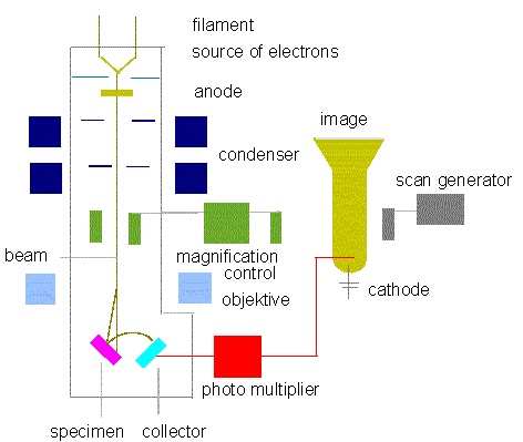
Block diagram of a typical SEM
(Redrawn from J. W. S. HEARLE, J. T. SPARROW, P. M. CROSS, 1972)
The properties of the light microscope as opposed to that of transmission and scanning electron microscopes are collected in a table .
Finally some outlines of new and further developments are given
.
- The high voltage electron microscope: it operates with an accelerating voltage of 700 - 3000 kV. Its power of resolution is greater, the preparation can be thicker, the strain on the preparation is smaller. But the enormous technical expenditure is disadvantageous. Only few gadgets exist. New results concerning botany have not been gained.
- The scanning transmission electron microscope (STEM): In this development of the SEM do the electrons pass through the preparation and the secondary radiation thus generated is used for image formation. Here, too, the expenditure is large, but it is still worthwhile, since large molecules like nucleic acids or proteins or molecular complexes like viruses can be depicted much better and gentler than with the TEM. No news for botany, though.
The interpretation of images gained with electron microscopy is increasingly done with computerized interpretation programs. But they are usually only suitable for the reconstruction of regularly recurring patterns and these, again, are found more often on a molecular level than on a cellular one.