Botany online 1996-2004. No further update, only historical document of botanical science!
Fluorescence
Microscopy
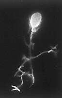 Fluorescence
microscopy is based on the fact that some molecules emit part of the
light absorbed by them as longer waves. A well-known example is the
red fluorescence of
chlorophyll. Since the Austrian teacher M. HAITINGER made his
examinations in the thirties of this 20th century it is known that a
number of so-called fluorochromes exist, with which microscopic
preparations can be stained so that they emit fluorescence
indirectly. In the following decades, a whole range of so-called
vital dyes were detected or developed, that -used in low
concentrations- could mark specific parts of living cells or of
tissues. They enabled researchers to follow the solute transport
within cells and tissues or to ascertain the pH of special
compartments, like for example the vacuole.
Fluorescence
microscopy is based on the fact that some molecules emit part of the
light absorbed by them as longer waves. A well-known example is the
red fluorescence of
chlorophyll. Since the Austrian teacher M. HAITINGER made his
examinations in the thirties of this 20th century it is known that a
number of so-called fluorochromes exist, with which microscopic
preparations can be stained so that they emit fluorescence
indirectly. In the following decades, a whole range of so-called
vital dyes were detected or developed, that -used in low
concentrations- could mark specific parts of living cells or of
tissues. They enabled researchers to follow the solute transport
within cells and tissues or to ascertain the pH of special
compartments, like for example the vacuole.
Since roughly twenty years fluorescence microscopy is flowering
again, on one hand, because the range of fluorochromes became much
broader and on the other hand, because completely new approaches like
indirect fluorescence could be used successfully. Additionally the
construction of microscopes was improved and highly efficient masks
were developed.
There are two ways in which fluorescence microscopes can be
constructed: as epifluorescence-
and as transmission microscopes.
The second is the older way of construction. Three components are
needed:
- A strong source of light, that emits mainly short
lightwaves. Mercury high pressure lights have proven
useful.
- The first barrier filter: This filter helps to shut off all
radiation other than the one that activates the specific dye.
It is placed behind the light source within the light cone.
Besides it is advantageous to work with a dark field
condenser.
- Second barrier filter: This filter is brought into the
light cone between objective and eyepiece. It lets through only
long wavelengths, that are caused by emission of the
preparation (so-called "secondary radiation").
In the last years the epifluorescence
microscope has replaced the transmission fluorescence
microscope more and more. But the transmission fluorescence
microscope is still better suited to only weakly magnifying
objectives (2.5 x, 6.3 x). With the epifluorescence the objective is
also a condenser and the stronger it is the more intensive radiation
can be used. The heart of epifluorescence is a construction within
the light cone between objective and eyepiece that serves to feed
activating radiation into the system and is constructed of first
barrier filter, beam-splitting mirror and second barrier filter.
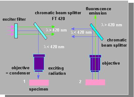
Excitation and fluorescence with chromatic beam splitters. Similar
to the interference filters these are specially coated mirrors
used under 45° to the illuminating beam. They reflect certain
spectral ranges, while others are completely transmitted. The
separating line between reflection and transmission may be set at
any point of the spectrum. 1.
Exciting radiation, 2.
Fluorescence emission. (Redrawn from diagram by CARL ZEISS).
© Peter v. Sengbusch - Impressum