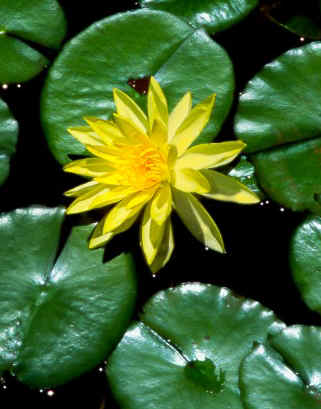
Leaves vary a great deal in terms of their general form and internal anatomy. Variations due to environmental interactions are most strongly expressed in leaves. For instance, the leaves of Ohi'a lehua (Metrosideros polymorpha) display several "morphotypes" which vary considerably is shape, thickness and pubescence. Most of the variation is due to phenotypic plasticity. This means that the environment interacts with the genes in the leaves to produce foliage variations. However, many species have genetically fixed adaptations which equip them to deal with stressful environments. Dry environments are called xeric while wet habitats are called hydric. Leaves can have xeromorphic or hydromorphic features. The term Mesomorphic is used to describe traits associated with leaves grown is moderately moist environments. It is possible for leaves to have a mixture of hydromorphic and xeromorphic traits. The leaf of water lily (Nymphia) is a good example. The term xeromorphic should probably be changed to stressomorphic because many kinds of stress can evoke the same traits as water stress. The term xeromorphic persists because these anatomical features were first documented for desert plants. Different traits are sometimes expressed at different periods in plant development.
 |
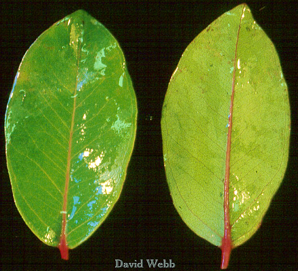 |
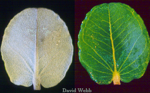
| Pubescent Ohi'a lehua Leaves | Glabrous Ohi'a lehua Leaves |
In the case of koa (Acacia
koa) the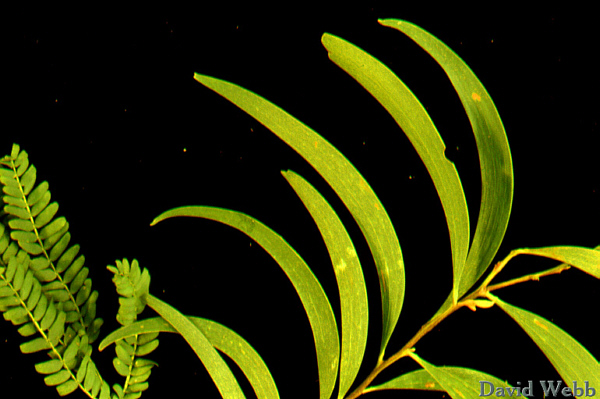
juvenile leaves are bipinnately compound and have tiny, delicate leaflets.
These are Mesomorphic. The mature foliage is a leaf-like appendage which is
actually the highly modified leaf rachis. The rachis is the central part of a
pinnately compound leaf to which the primary leaflets are attached. This kind of structure
is called a phyllode. The mature "leaf" has xeromorphic
adaptations.
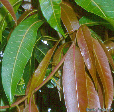
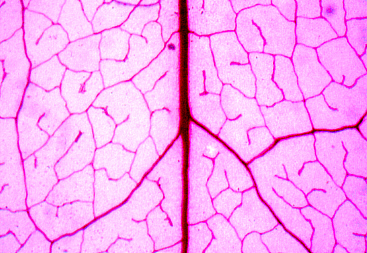
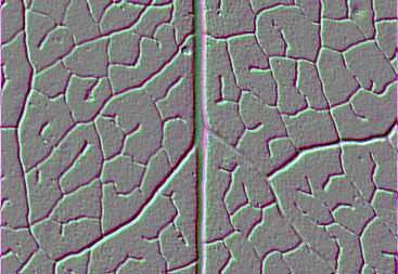
Dicot leaves of tend to have either pinnate or palmate Organization. Examine the Glabrous Ohi'a lehua leaves above to see an example of Pinnate Venation.
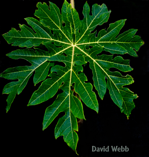
See the examples below and sort out which leaves are pinnate or palmate.
 Leaf with Pinnate Venation |
|
|
|
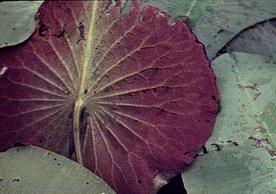
Dicot leaves generally have a large midrib which gives rise to the first order of lateral veins. Branching of the lateral veins occurs with each branch being progressively smaller. The ultimate branches surround small areas of the leaf (areoles). The overall pattern resembles a net. Thus, the venation of dicots is called net or reticulate. Cross sections of leaves with reticulate venation show several size classes of veins in various planes of section. Some veins will be perfect cross sections while others will be oblique or parallel to the plane of section.
Monocot leaves, like those of Ti and Sugarcane, tend to be elongated. They generally exhibit striate venation because the major veins run parallel to one another at some point in the leaf. The veins converge towards the apex and form a unified closed system. Many veins can be perpendicular to the plane of a cross section. Consequently, they produce a relatively orderly vein pattern compared to dicots. However, a close inspection reveals that lateral connections exist between parallel veins. These are called commissural bundles. Furthermore, some monocots have net venation which is difficult to separate from dicots. The bottom line is that dicots and monocots have reticulate venation, however striate venation is different from the reticulate venation found in most dicots.
|
|
|
|
We will use cross sections to study leaf anatomy. However, we will have demos of one to several paradermal sections. These are cut parallel to the surface (approximately) and reveal details of the anatomy from a different perspective. In reality paradermal sections are not perfect but cut through the leaf at an oblique angle. This allows you to start with the upper epidermis and scan through the remaining layers.
Study cross and paradermal sections
of Syringa (lilac). This has "textbook" leaf anatomy.
Start with the upper epidermis and work through a cross section from top to
bottom.
|
|
Midrib (left) and blade (above) of a "textbook" leaf. Identify the various layers. |
|
Epidermis
Is there an obvious cuticle?
Is it uniseriate or multiseriate?
Are there noticeable differences between the upper (adaxial) or lower (abaxial) Epidermis?
Are there any differences between epidermal cells associated with veins, compared to epidermal cells situated above and below photosynthetic tissues?
On which surface(s) can you find stomata?
Are trichomes present?
Mesophyll
Note the differences between the Palisade and Spongy layers.
What is the degree of intercellular spaces among the Spongy and Palisade?
Is there any obvious functional significance for these differences?
Vascular Tissues
Note the different sizes of the veins in the midrib and lamina (blade)
Are the vascular bundles collateral?
Are both Vascular Tissues present in both
Are Bundle Sheaths visible?
Where do you find the most supporting tissues (Collenchyma or Sclerenchyma)?
Compare fresh sections of Ohi'a lehua and Koa with the "Textbook" leaf.View unstained & then with Polarizers.
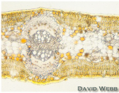
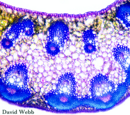
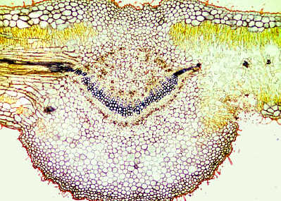
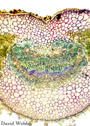 Midrib of Ohi'a lehua stained with Toluidine Blue |
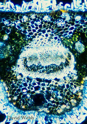 Unstained Ohi'a lehua Midrib seen with Polarized Light. |
|
|
|
|
|
|
Young Koa with Juvenile & Mature Foliage |
|
 Juvenile Koa Leaf |
 Juvenile Koa Leaf |
 Koa Phyllodes and denuded Rachis from a Juvenile Leaf |
|
 Cleared Koa Leaflet |
 Cleared Phyllode |
||
|
|
|
|
Monocot Leaves
Study commercial slides of Sugarcane (Saccharum officinarum)
Epidermis
Is there an obvious cuticle?
Is it uniseriate or multiseriate?
Are Bulliform Cells present? What is
their function(s)?
Are there noticeable differences between the upper (adaxial) or lower (abaxial) Epidermis?
Are there any differences between
epidermal cells associated with veins, compared to epidermal cells situated above and
below photosynthetic tissues?
On which surface(s) can you find stomata?
Are trichomes present?
Mesophyll
Can you find Palisade and Spongy layers?
If NOT, what term is used to designate the arrangement of photosynthetic tissues in leaves like this?
What specific type of photosynthesis does this plant possess?
Where do you find the intercellular spaces and stomata in this leaf?
Is there any obvious functional significance for these differences?
Vascular Tissues
Can you find a midrib ?
Note the different sizes of the veins in the lamina (blade). Might they have different functions based on their size and complexity?
Are the vascular bundles collateral?
Are both vascular tissues present in all of the veins in the same proportions??
Are bundle sheaths visible? Do they have extensions? Are they the same for major and minor veins?
Where do you find the most supporting tissues (Collenchyma or Sclerenchyma)?
Examine fresh sections. View unstained then stain with Toluidine Blue.
Observe demo of a leaf with a fluorescence microscope & note the distribution of Chlorenchyma.
Examine intact leaves
of Cordyline with a dissecting scope or hand-lens.
Note the orientation of the veins.
Place a 2 x 2 cm piece on a microscope
slide and place
this onto the microscope stage.
Use the 4X objective and swing up the
high power condenser lens.
Turn the illumination knob to maximum!
Look for the Commissural Bundles!
Observe cross sections
from fresh leaves and stain with Toluidine Blue.
Observe the demo of a Loulu Palm Leaf & ID the Venation.
Midribs
The anatomy of midribs can be complex for both Monocots and Dicots. There my be one to several major bundles with Dicots. Many similarly sized vascular bundles constitute the "Midrib" of monocots.
Examine fresh sections and a demo showing the midrib of Ti.
Do the same for Ohi'a lehua.
Examine the midribs in demo slides of other species.
|
|
Examine demos of cleared Dicot and Monocot Leaves. These can include
Teak (Tectona), Tamarind (Tamarindus), Ti (Cordyline), Loulu Palm (Pritchardia), Anthurium, Kalo/Taro (Cocolasia)
Determine which have Reticulate and those that have Striate venation.
Wherever possible compare fresh leaves with the cleared leaves.
|
|
|
|
Cleared Leaves with Striate or Reticulate Venation
|
|
|
|
|
|
| Dichotomous Venation: We focus on seed plants in this course. However, there are many terrestrial plants that do not produce seeds. Ferns are the most familiar of these. Many species have dichotomous venation. Dicho means two. Tomo refers to cut. Thus dichotomous means to cut in half. Dichotomous venation occurs when veins branch at the tip such that two branches result. These will do the same and establish a 2 x 2 x 2 ...... vein system. The ultimate veins may end blindly at the margins of the leaf (open dichotomous) or they may unite with other veins and form a closed dichotomous system. Dichotomous venation is found in Cycads and some conifers like Agathis which are seed plants but it is more common in "ancestral" or "primitive" vascular plants, like ferns. |  Fern with Open Dichotomous Venation
|
Examine the venation of Zamia or Agathis leaves.
Petioles
Leaves are generally attached to the stem by petioles. These often have a stem-like anatomy. However, the vascular tissues usually have an unusual arrangement. These patterns range from the simple to the very complex.
Many monocots do not have separate petioles but are attached to the stem by leaf-like sheaths which may encircle and embrace the stem.
Examine commercial
cross sections of
Cycas (Gymnosperm) Camellia and Horse Chestnut (Aesculus hippocastanum)
petioles.
Examine fresh sections of various petioles from local plants, including. Ohi'a Lehua & Koa
Many monocots do not have petioles but have a sheath that connects the blade (lamina) to the stem.
Compare cross sections of Ki/Ti to sections from its midrib and lamina.
|
 Horse Chestnut Petiole |
|
Stipules are leaf like structures which develop from leaf primordia early in development. They usually are sessile and ephemeral. They may protect young, emerging leaves. In some cases the stipules become protective thorns.
Examine the leaf-like stipules on Hau
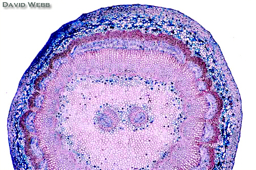
Gymnosperm Leaves
Most gymnosperms do NOT produce leaves
with wide blades. Their  leaves resemble needles or
scales. In the case of Pinus monophylla (one leaf) the leaves are cylindrical. These
shapes are xeromorphic adaptations because they reduce the amount of surface area
available for evaporation. They have many other xeromorphic adaptations which include a
thick cuticle, sclerified epidermal cells, sunken stomata, a sclerotic hypodermis, tightly
packed mesophyll, an endodermis, few to no lateral veins and centrally located vascular
tissues. There is also a great deal of lignification. A specialized Transfusion Tissue
often surrounds the vascular bundle. It contains tracheids and performs the function of
lateral veins.
leaves resemble needles or
scales. In the case of Pinus monophylla (one leaf) the leaves are cylindrical. These
shapes are xeromorphic adaptations because they reduce the amount of surface area
available for evaporation. They have many other xeromorphic adaptations which include a
thick cuticle, sclerified epidermal cells, sunken stomata, a sclerotic hypodermis, tightly
packed mesophyll, an endodermis, few to no lateral veins and centrally located vascular
tissues. There is also a great deal of lignification. A specialized Transfusion Tissue
often surrounds the vascular bundle. It contains tracheids and performs the function of
lateral veins.
Examine commercial slides of Pinus leaves (needs) and locate the anatomical features stated above.
Examine fresh sections of Norfolk Island Pine (Araucaria).
Use your polarizers on unstained sections.
Stain several sections with phloroglucinol to locate lignified cell walls.
Locate the Endodermis! What might its function be?
|
|
|
|
Variation in Leaf Structure
Xeromorphic & Hydromorphic Leaf Traits
Hydromorphic Traits |
Xeromorphic Traits |
| Blades broad, flat, thick (high surface/volume ratio) |
Blades
narrow, thick, even cylindrical (low surface/ volume ratio) |
| Trichomes few to none | Trichomes present |
| Thin Cuticle | Thick Cuticle |
| Normal Epidermis | Sclerotic Epidermis |
| No Hypodermis | Hypodermis Present |
| Adaxial & Abaxial Stomata | Abaxial Stomata & Sunken Stomata or Stomatal Crypts |
| Relatively low level of Palisade Parenchyma | Relatively high proportion of Palisade Parenchyma |
| Bifacial | Unifacial |
| Aerenchyma Prominent | Little intercellular spaces |
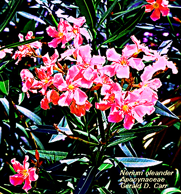
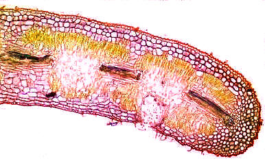
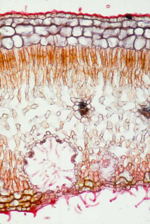
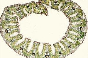
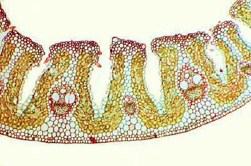
We will use Nerium oleander (dicot) and Amophilia (monocot) as examples of leaves with Xeromorphic Traits.
Obtain commercial slides.
Orient the leaves so that the upper (adaxial) surface is correctly positioned on your microscope.
Start with the Upper (Adaxial) Epidermis and note whether or not Xeromorphic traits are evident as you work your way through the leaf.
YES means that a Xeromorphic trait is Present.
|
|
|
|
| Traits of Nerium oleander | No | Yes |
| Overall Shape | ||
| Abundant Trichomes | ||
| Thick Cuticle | ||
| Adaxial Stomata ABSENT | ||
| Sunken Stomata | ||
| Hypodermis (H) or Multiple Epidermis (ME) | ||
| Abundant Palisade (Unifacial) | ||
| Little to No Spongy Mesophyll | ||
| Abundant Crystals | ||
| Abundant Sclerenchyma associated with the Vascular Bundles | ||
| Sclerenchyma
Elsewhere (E = Epidermis; M = Mesophyll; H = Hypodermis) |
||
| Kranz Anatomy | ||
| Bundle Sheath Extensions | ||
| Lower Hypodermis (H) or Multiple Epidermis (ME) | ||
| Sclerefication of the Hypodermis | ||
| Sunken Stomata | ||
| Stomatal Crypts | ||
| Abundant Trichomes | ||
| Other |
|
|
| Traits of Amophila | No |
Yes |
| Overall Shape | ||
| Abundant Trichomes | ||
| Thick Cuticle | ||
| Adaxial Stomata ABSENT | ||
| Sunken Stomata | ||
| Hypodermis (H) or Multiple Epidermis (ME) | ||
| Abundant Palisade (Unifacial) | ||
| Little to No Spongy Mesophyll | ||
| Abundant Crystals | ||
| Abundant Sclerenchyma associated with the Vascular Bundles | ||
| Sclerenchyma Elsewhere (E = Epidermis; M = Mesophyll; H = Hypodermis) |
||
| Kranz Anatomy | ||
| Bundle Sheath Extensions | ||
| Lower Hypodermis (H) or Multiple Epidermis (ME) | ||
| Sclerefication of the Hypodermis | ||
| Sunken Stomata | ||
| Stomatal Crypts | ||
| Abundant Trichomes | ||
| Other |
|
|
 Nymphaea Leaf Cross Section |
| Traits of Nymphaea | No |
Yes |
| Overall Shape | ||
| Abundant Trichomes | ||
| Thick Cuticle | ||
| Adaxial Stomata ABSENT | ||
| Sunken Stomata | ||
| Hypodermis (H) or Multiple Epidermis (ME) | ||
| Abundant Palisade (Unifacial) | ||
| Little to No Spongy Mesophyll | ||
| Aerenchyma | ||
| Abundant Crystals | ||
| Abundant Sclerenchyma associated with the Vascular Bundles | ||
| Sclerenchyma Elsewhere (E = Epidermis; M = Mesophyll; H = Hypodermis) |
||
| Kranz Anatomy | ||
| Bundle Sheath Extensions | ||
| Lower Hypodermis (H) or Multiple Epidermis (ME) | ||
| Sclerefication of the Hypodermis | ||
| Sunken Stomata | ||
| Stomatal Crypts | ||
| Abundant Trichomes | ||
| Other |
Fill out our favorite Table for Pinus leaves!
| Traits of Pinus | No |
Yes |
| Overall Shape | ||
| Abundant Trichomes | ||
| Thick Cuticle | ||
| Adaxial Stomata ABSENT | ||
| Sunken Stomata | ||
| Hypodermis (H) or Multiple Epidermis (ME) | ||
| Abundant Palisade (Unifacial) | ||
| Little to No Spongy Mesophyll | ||
| Abundant Crystals | ||
| Abundant Sclerenchyma associated with the Vascular Bundles | ||
| Sclerenchyma Elsewhere (E = Epidermis; M = Mesophyll; H = Hypodermis) |
||
| Kranz Anatomy | ||
| Bundle Sheath Extensions | ||
| Lower Hypodermis (H) or Multiple Epidermis (ME) | ||
| Sclerefication of the Hypodermis | ||
| Sunken Stomata | ||
| Stomatal Crypts | ||
| Abundant Trichomes | ||
| Other |
![]()