Water Hyacinth (Eichornia crassipes) is an
aquatic plant which floats on lakes and slow-moving streams 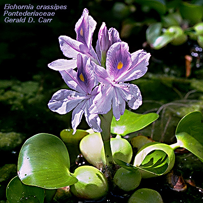 of
rivers. It is incredibly beautiful but it can be an environmental
nightmare! It grows at an incredible rate and can be a serious environmental pest.
It doesen't help that its natural herbivor, the Manatee has been driven to the brink of
extinction.
of
rivers. It is incredibly beautiful but it can be an environmental
nightmare! It grows at an incredible rate and can be a serious environmental pest.
It doesen't help that its natural herbivor, the Manatee has been driven to the brink of
extinction.
It quickly becomes a complete mat which crowds out other floating aquatic plants like Lemna or Azolla.
It also intercepts all of the incident illumination that phytoplankton might use otherwise.
The dead biomass produced by Eichornia accumulates on the bottom and is decomposed by by microorganisms.
Because there is a tremendous amount of dead biomass, the environment becomes anaerobic as the available oxygen is consumed by the decomposers.
This is a viscous circle which further reduces the ability of the ecosystem to support other species.
The roots are totally submerged and have some adaptations associated with growth in a Hydric environment. Such traits are called Hydromorphic.
 |
| Cross-section of Water Hyacinth root stained with Toluidine Blue. |
The tissues starting from the outside to the Inside are

Epidermis
Ground Tissue (Outer Cortex)
Ground Tissue (Cortex)
Aerenchyma
Aerenchyma is typically found in submerged organs.
It is a Hydromorphic Trait.
Ground Tissue (Inner Cortex)
Endodermis
Pericycle
Phloem
Xylem
Ground Tissue (Stele)
The Pericycle (around the circle) is parenchyma located immediately inside the Endodermis. It may be one to several cell layers thick. It tends to be several layers thick in monocot roots. It is one source of Lateral Root initiation.
To locate the Endodermis, find a file of cells from the Inner Cortex and follow it as far as you can towards the center of the root. This unicellular layer located by this method is the Endodermis. It will be hard to see the Casparian Strips.
|
| The Pericycle is hard to see. It appears to be one cell thick and appears incomplete because it is collapsed by cell expansion in the stele. |
|
The large Vessel Members (VMs) of
the Metaxylem are easy to find. Locate the other Vessel
Members by looking immediately towards the the OUTSIDE of the
largest VMs. These VMs will have smaller diameters. The smallest
represent Protoxylem. This is EXARCH
development. The direction of xylem maturation is from the outside (Ex) towards the
Inside. |
The Phloem alternates
with the xylem. You can see an enlarged Sieve Tube Member in
each. The phloem cell walls will be pink because they lack lignin. |
| The
center of the stele is composed of Parenchyma. This is typical of monocots but virtually
absent in Dicots. These cells may be sclerified in terrestrial monocot roots. Absence of Sclerenchyma is another Hydromorphic Trait. |
Two illustrations of corn roots are found below. Find the tissue layers and compare with Water Hyacinth. Note the presence of a few Root Hairs. Absence of Root Hairs is a Hydromorphic Traits. |
|
|
|
| Embossed Images of a Corn Root | |


