|
|
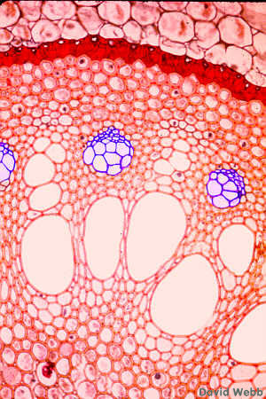
| Vascular Tissues in a Corn Root - The cells with the largest Diameters are Vessel Members. | Interface of the Stele & the Cortex-Follow the Cortical Parenchyma from the top of the image to the first cells which have thick, red-stained walls. These form the Endodermis. |
The Endodermis usually develops exceedingly thick secondary walls at levels in the root which are no longer absorbing water. This obscures the Casparian Strips but makes the Endodermis more obvious.
The organization of Monocot roots like Sugarcane is similar to that found in dicots like Ranunculus. However, tracheary elements may be absent from the center of the root. There are usually more xylem arms in monocots. The number of arms is indicated by the following terms.

| Two = Diarch |
| Three = Triarch |
| Four = Tetrarch |
| Five = Pentarch |
| Six = Hexarch |
| More than five = Polyarch |
Monocots are usually Polyarch. Bundles of Phloem alternate with the Xylem arms. The xylem contains many lignified cells in older portions of the root. The phloem stands out because its cells have thin, unlignified cell walls.
|
Cross-section of Clintonia (monocot) Root. Count the number of xylem arms. |
|
|
Cross-section of Smilax
(monocot) Root. The tracheary elements of the Xylem are the cells with the largest
diameters. Count the number of xylem arms.
The cell pattern is distorted by the extreme enlargement of the Vessel Members. |
Smilax root at higher magnification. Locate the Xylem and Phloem as well as the Endodermis, Cortex and Epidermis.
|