"Simple"
Tisues - Parenchyma - Collenchyma & Sclerenchyma

This lab is designed to give you information on the primary
nonvascular tissues. These are relatively simple compared to xylem and phloem.
However, we will see that there is a considerable amount of variation within these
tissues. In addition, you will observe the major components of the protoplast that are
visible with the light microscope.
Study cell shape, contents, and wall structure, the relation of
cells to one another for intact tissues, the presence of intercellular
connections via pits, and the presence or absence of intercellular spaces. The cell
walls and air spaces constitute the Apoplast. The Plasmalemma and all
within it constitute the Symplast. These are Extremely Important concepts,
which must be appreciated to understand Plant Physiology!
Within the Symplast,
look for the cytoplasm, nuclei, chloroplasts, other plastids, crystals, and vacuoles
colored with anthocyanins. Use polarizing filters to locate starch grains and
crystals. Also use polarizers and stains to study cell wall organization and
composition.
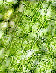
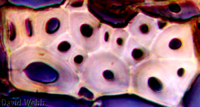
PARENCHYMA
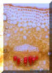
Cortex of Pereskia stem: 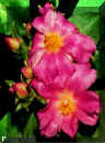
Observe
Parenchyma
consists of relatively large, thin-walled cells. The cells are arranged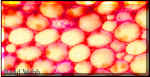 loosely, that is, there are intercellular spaces among them. The protoplasts of
these cells contain chloroplasts. Some of these cells may have amyloplasts
and crystals. Pereskia is a member of the cactus family. It has spines but it also
has normal leaves. Its flowers are extremely beautiful like those of most cactaceae.
loosely, that is, there are intercellular spaces among them. The protoplasts of
these cells contain chloroplasts. Some of these cells may have amyloplasts
and crystals. Pereskia is a member of the cactus family. It has spines but it also
has normal leaves. Its flowers are extremely beautiful like those of most cactaceae.
Stain
Young leaf of Elodea.
Mount
Use
Observe demo with phase contrast optics to study the cytoplasm.
Observe
Macerated Pith of Begonia (prepared by boiling in dilute KOH): Note
numerous faces of individual cells. What term is used to describe cells which have
this shape?


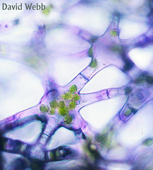
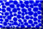
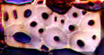
Aerenchyma & Stellate Parenchyma
A
strikingly different shape of parenchyma cells is illustrated by stellate parenchyma.
These are branched and adjacent cells are connected with each other by means of the
branches. Parenchyma composed of branched cells is highly lacunose; that is, it has a
large volume of intercellular space. The spongy layer in leaves has branched cells with
large intercellular spaces. The term Aerenchyma is often used to describe parenchyma,
which has large air spaces.
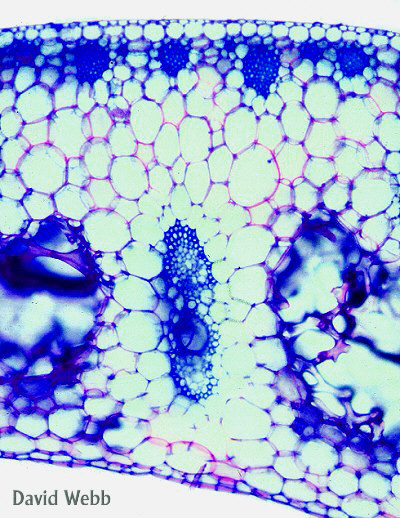
Locate Stellate
Parenchyma cells in petioles and midribs of Canna (ali’iope)
leaves. Cut hand sections and examine with a dissecting scope before observing with a
compound microscope. Do these have 3D branching?
Examine
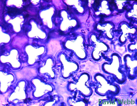
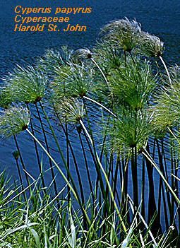
the Parenchyma in Papyrus (Cyperus papyrus) stems by making transverse
sections. Find the Aerenchyma with a dissecting scope and examine with a compound
microscope. What is the shape of the individual cells which comprise the Aerenchyma? Are
they branched in 2D or 3D?
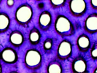
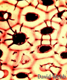
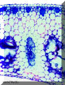
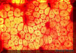
Cyperus papyrus stem stained with IKI
|
Aerenchyma in Cyperus javanicus |
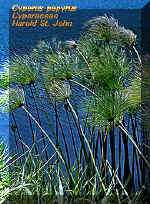
Cyperus papyrus |
Cyperus laevigatus
(makaloa) |
Aerenchyma
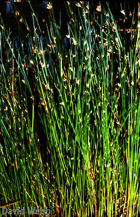
can be found in the stems of other members of the Cyperaceae, like C. laevigatus
(makaloa). Makaloa stems are smooth and resilient. The Aerenchyma
is like foam rubber on a microscopic scale. Makaloa stems
were used to make fine sleeping mats by ancient Hawaiians and the qualities of their
Aerenchyma contributed to their utility.
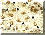
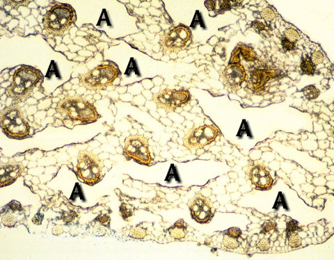
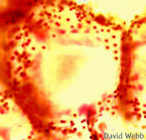
Sample from a makaloa mat: Note the Air Spaces (A)
|
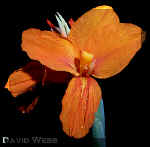
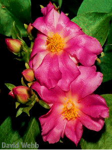
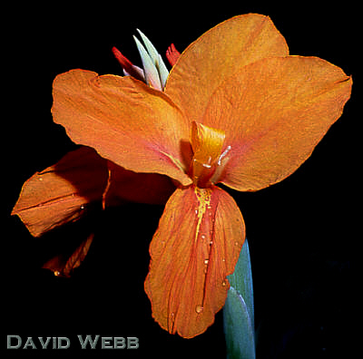
Canna Flowers
|
Pineapple
leaves
also contain stellate parenchyma. What functions are suggested by
the 3-dimensional shape of these cells?
Canna Petiole Cross Section
|
Stellate Parenchyma Cells
|
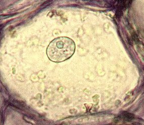
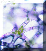
Endosperm Cells:
The parenchyma cells you have
examined thus far have relatively thin walls, but there are also thick-walled
parenchyma cells.
Examine
the demonstration slide of persimmon or palm (niu)
endosperm. This material will also show fine lines traversing the thick walls from cell lumen
to cell lumen. These lines are pits, which connect the symplast of adjacent
cells.

Persimmon Endosperm
|
Endosperm with Large Pits
|
You have
already observed cytoplasm and chloroplasts. Other protoplast components
include several more types of plastids, vacuoles, and various kinds of crystals.
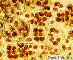
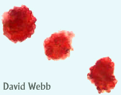
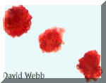
Chromoplasts
and Pigment Bodies. They may be yellow, red, and orange colored plastids and
similarly colored crystal-like bodies. The latter are called pigment bodies
because there is some question whether they may be classified as plastids.
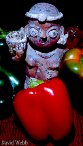
Observe
chromoplasts and pigment bodies in free-hand sections of bell pepper
fruits, various flower petals, and the root of carrot. Chloroplasts
are Chromoplasts, as well!
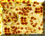
Chromoplasts from Red Pepper (Capsicum)
|

Chromoplasts from Flower Petals
|

Red Bell Pepper Fruit |

The color of Alamanda Flowers is due to Chromoplasts |
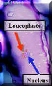
Leucoplasts
are mysterious and difficult to
demonstrate without special techniques.
Observe
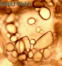
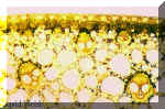
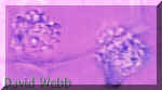
Amyloplasts
are filled with starch, which sometimes occupies the entire organelle.
They are also regarded as Leucoplasts because they lack color.
Observe
thin free-hand sections of Papyrus and stain
with IKI. We will have a demo of potato amyloplasts.
Observe
an unstained specimen and use the polarizers.
View a stained
slide and then use the polarizers.
|
Leucoplasts clustered around the Nucleus of a
Parenchyma Cell stained with Toluidine Blue |
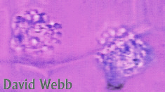
Unstained potato Amyloplasts
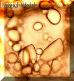
|
Commercial slide of Potato Amyloplasts
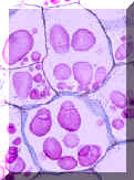
|
Amyloplasts from Canna seen with normal illumination |
Amyloplasts from Canna seen with crossed Polarizers |
Amyloplasts stained with IKI
|
"Statoliths" are amyloplasts, which
contain many large multifaceted starch grains, similar to those above. Their
function may be related to gravity perception.
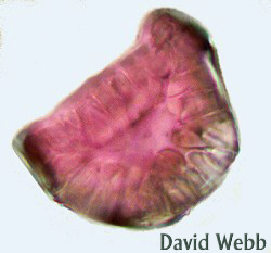
Non-cytoplasmic Contents
Make
slides of Rhoeo or Zebrina epidermis (see above for
Leucoplasts). These demonstrate vacuoles, which contain
anthocyanin.
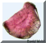
The
pigmentation in many flower petals, like Erithrina (wiliwili), is also
contained in vacuoles. This is best observed by looking at fresh cross sections of
the petals. How can you tell if the color is due to chromoplasts or vacuolar
pigments?
Anthocyanins in a surface view of Epidermal Cells
|

Zebrina Leaves: The Anthocyanins are on the lower surface of the Leaves. I
wonder what they are doing down there????
|
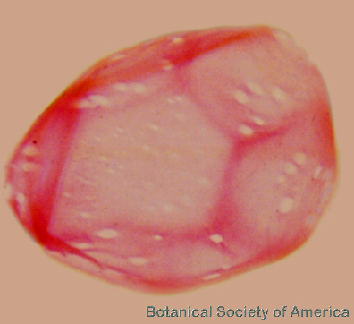
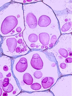
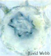
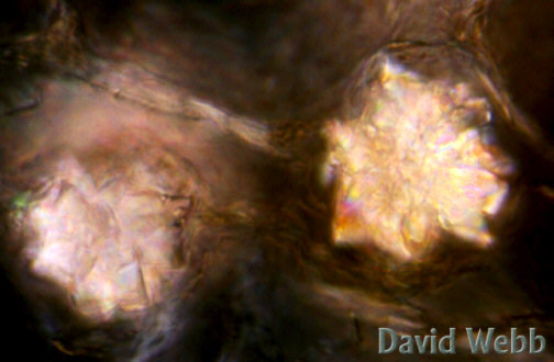
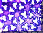
Crystals are vacuolar
in nature.
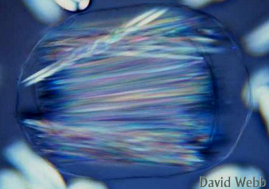
Observe Raphides
Druse Crystal seen with Bright Field Illumination
|
Druse Crystals seen with Crossed Polarizers.
|
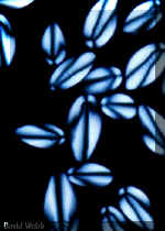
Raphides in an isolated Vacuole seen with crossed Polarizers
|
Raphides on the Loose!!!!
|
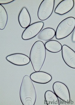
Observe Prismatic
 outer, dry scales of Allium cepa (onion)
bulbs. These scales have been soaked in alcohol to remove the air.
outer, dry scales of Allium cepa (onion)
bulbs. These scales have been soaked in alcohol to remove the air.
Try your
polarizers on these!!!!!
The function of these crystals is relatively uncertain.
They
seem to be more abundant in plants, which grow in arid and xeric environments. They are
all composed of Calcium oxalate, which causes epithelial cells to swell.
Consequently, they should deter herbivory.
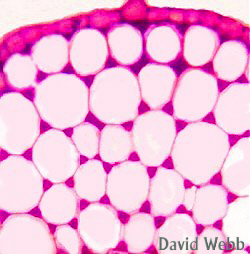
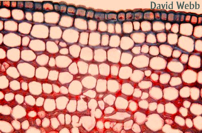
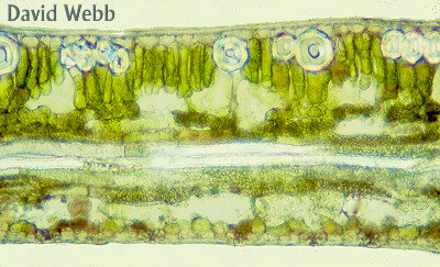
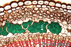
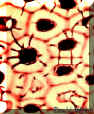
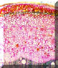
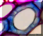
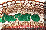
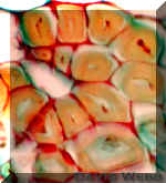
COLLENCHYMA
Collenchyma
is closely related to parenchyma. However, the plastids are not well differentiated
in collenchyma while they are well differentiated and obvious in parenchyma. Collenchyma
always occurs just beneath the epidermis, while parenchyma occurs throughout the
plant. Collenchyma cell walls are unevenly thickened. When the
thickening occurs at the corners where cells are joined it is called angular. Lamellar
collenchyma has thickenings on their tangential walls, which are parallel with
the surface. Lignin is usually not present
in collenchyma.
Locate
Collenchyma in hand sections of Widelia stem, Celery
or Water Lily (Nymphia) petioles. Determine cell shape by
observing cross sections and a demo of a longitudinal section.
Mount fresh sections in water. After examining them, stain with Toluidine Blue
and then examine again. What does the pink color of the cell walls indicate?
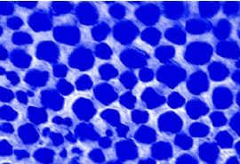
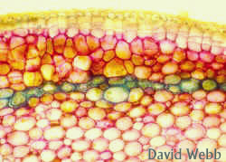
Observe
Unstained Collenchyma in Widelia Stem
|

Collenchyma stained with Toluidine Blue
|
Unstained Collenchyma in Celery
|

Unstained Collenchyma in Celery
|
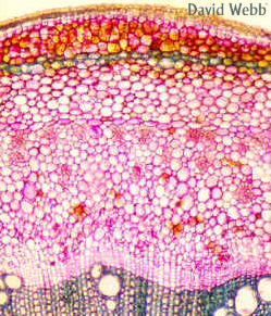
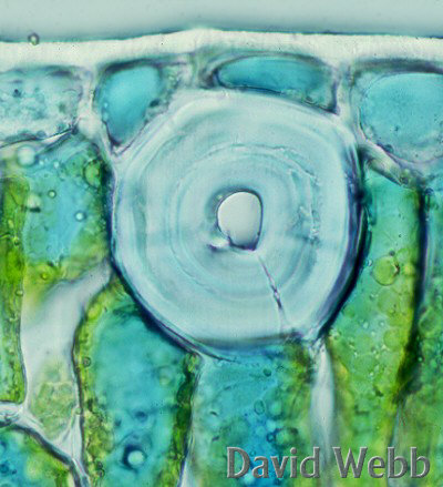
Observe
(prepared slides) of Sambucus stems.
The thickenings are chiefly on the tangential walls. Tangential
in these case means walls oreiented parallel to the surface of the
structure. What type of Collenchyma is this?
thickenings are chiefly on the tangential walls. Tangential
in these case means walls oreiented parallel to the surface of the
structure. What type of Collenchyma is this?
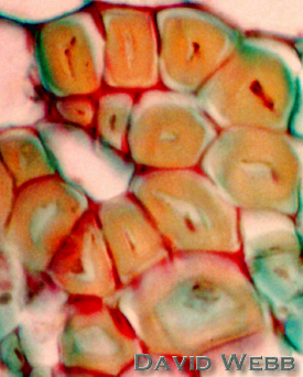
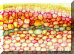
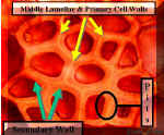
Sclerenchyma
The
distinction between parenchyma, collenchyma and sclerenchyma is largely based on the wall
structure. Parenchyma cell walls are usually thin and primary while in sclerenchyma
a secondary wall is formed on the inner side of the primary wall. Secondary
walls are those, which develop after a cell, has ceased to enlarge. Collenchyma
cells have secondary wall thickenings but these are uneven in their
distribution. Furthermore, the cellulose fibrils in Collenchyma are not as highly
organized or tightly bound as in Sclerenchyma. Finally, Sclerenchyma cells can be
found in many locations throughout the plant body but Collenchyma are always just
beneath the Epidermis.
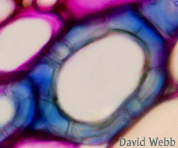
Sclerenchyma
cells are usually classified into sclereids or
fibers on the basis of form as well as the abundance and type of
pitting.
Sclereids
are generally shorter than fibers and
their walls show more abundant pitting. The pits are often branched (ramiform).
Walls of sclerenchyma cells are usually lignified and, therefore, stain red
with safranin or phloroglucinol-hydrochloric acid. They often show concentric laminations,
which indicate different periods of wall synthesis. Sclereids vary in shape
and occur in all parts of the plant.
Fibers
tend to be highly elongated cells with tapering
ends, and they often occur in bundles. There are few pits in the walls of
fibers. The pits, when present, are usually simple and unbranched.
In studying
Sclerenchyma observe their (1) overall shape;
(2) wall structure; (3) pits; (4) staining reactions to Phloroglucinol
& Toluidine Blue; (5) appearance with crossed polarizers.
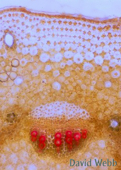
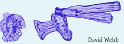
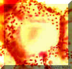
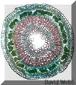
Sclereids
from the stem of Hoya (wax plant). Sclereids
occur between the cortex and the vascular region, and in the pith.
They resemble parenchyma cells in shape but have thick walls. A comparison of the
sclereids with the adjacent parenchyma cells illustrates the two extremes in the variation
of plant cell walls
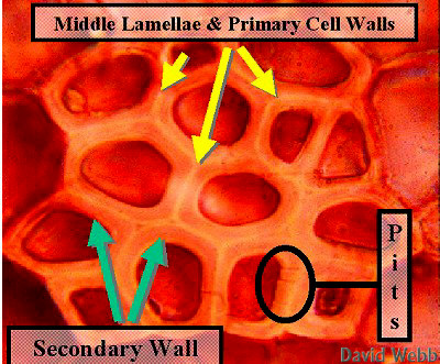
transverse sections with Toluidine Blue. Use older stems for lignified
sclereids. The parenchyma cells have thin primary, nonlignified walls. The
sclereids have a thick lignified secondary wall deposited inside the thin
primary wall. The secondary wall obscures the primary wall and shows concentric
lamination because it is deposited in successive layers. It also shows prominent
canal-like pits. To observe details of the pits, focus up and down while
examining them. The primary walls of adjacent sclereids, and the middle lamella are
tightly joined and obscured by lignin deposition. Lignin makes the cell walls very
strong and waterproof.
|
|
| Cross Section of Hoya
Stem stained with Toluidine Blue: The sclereids occur in a unicellular band in the outer
part of the stem. |
Solitary Sclereid on Hoya Stem stained with Toluidine Blue
|
Various Wall Layers in Sclerenchyma |
Examine
Mount
Examine
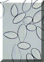
Elongated Sclerids in Onion Bulb Scale
|
Cross section of Podocarpus Leaf
|

Astrosclerids from Nymphaea Leaf
|
Sclereid from Podocarpus Leaf
|
Partly Dissected leaf showing Trichosclereids
|

Trichosclereids from Olive Leaf
|
Observe
Cluster of Brachysclereids
|
Solitary Brachysclereid
|
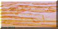
Fibers
Like
sclereids, fibers may be found in various parts of the plant. Fibers are
particularly common near the phloem (phloem or bast fibers) and the
xylem (xylem fibers). In monocots fibers often enclose vascular bundles (fibrovascular
bundles) or appear as strands that are independent of vascular tissues.
The best
commercial fibers are usually associated with the phloem. This includes hau
(Hibiscus tiliaceus) and wauke (Broussonetia papyrifera)
plus Cannabis. Coarse fibers can be obtained from monocot leaves like uki
uki grass (Dianella sandwichensis) and Agave. Agave was grown in Hawaii but was uneconomical. Some of
these plants have escaped cultivation and can be found in nature. They are slow
growing but once established, they may be difficult to eradicate. This could present a
problem for native species if they can't compete with Agave.
Mount
The phloem fibers of this plant were used by ancient Hawaiians for
making rope.
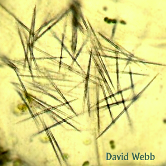
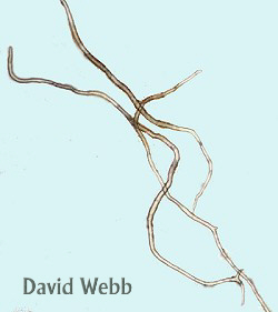
Unstained hau fibers from an Hawaiian artifact
|
Hau fibers stained with Phloroglucinol & viewed with crossed polarizers
|
Using prepared
slides, compare fibers Linen (Linum) and Hemp (Cannabis).
Note the fact that the linen fibers do not stain for lignin. Lignin makes the
fibers brittle and it discolors them as well.
|
Cross sections of flax stem that show the
phloem or "bast" fibers which are green. In this case the green color indicates
the absence of Lignin. |
Cannabis stem cross section
|
Highly Magnified Fibers
|
Fiber from Oak wood

Our Favorite Sclereid!!!!!