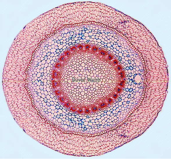
EPIDERMIS
The Epidermis is in direct contact
with the external environment. It contains many important adaptations which
allow plants to survive & reproduce on land. We will observe the most general
adaptations as well as some exotic ones. The functions of many types of epidermal cells
are well known but there are some specialized cells with unknown functions. The epidermis
is important in both vegetative and reproductive organs. It is treated here
in a broad sense as the superficial layer (or rarely layers) on all differentiated parts
of the plant in the primary state of growth. During secondary growth the
epidermis is often replaced by Periderm.
Many features of the epidermis can be seen in
whole mounts at low magnification with the compound microscope.Mount a piece of a Coleus leaf on a microscope slide.
Be sure to have the 4X objective in place.
Use tape to secure the leaf at each end. Do NOT
 cover the leaf with tape!!!
Place the slide on the microscope stage.
Move it so that the leaf is under the objective.
Be sure that the condenser iris is wide open.
Flip UP the swinging lens and turn the illuminator up. You may need to use maximal illumination.
Observe!!!!!
Note the differences between the upper and lower surfaces as well as the veins vs the lamina.You may be able to use the 10X objective but be careful NOT to bring it into contact with the leaf. THICK paradermal (parallel to the surface) sections or peels may be needed to observe epidermis from stems because of their thickness.
I learned to do this by accident!!!!!!!!
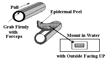 Epidermal
Peels: Epidermal features can also be captured by making peels. In many
plants, particularly succulent ones, the epidermis may be easily stripped from fresh
leaves. Such epidermal peels are useful for the study of the shape of epidermal cells and
their arrangement as well as the distribution and structure of stomata. To make epidermal
peels you should do the following.
Epidermal
Peels: Epidermal features can also be captured by making peels. In many
plants, particularly succulent ones, the epidermis may be easily stripped from fresh
leaves. Such epidermal peels are useful for the study of the shape of epidermal cells and
their arrangement as well as the distribution and structure of stomata. To make epidermal
peels you should do the following.
Make a clean cut at one end of the structure.
Use fine forceps to clamp down on a thick spot along the cut which includes the epidermis as the top layer.
Pull the forceps forward
Place this face up on a microscope slide and crop it so that some of the thin strip is retained.
Add water and observe!!!
Paradermal Sections: If the epidermis is not easily peeled, thin sections cut parallel with the leaf surface (paradermal sections) may be used. The latter will be demonstrated but are hard to do.
Epidermal Windows: There is a method of looking at the epidermis of leaves that I call the window method.
Place a Band-Aid on your index finger.
Roll the leaf over the Band-Aid.
Use a razorblade to scrape away the overlying tissues without cutting all the way through.
A window of translucent tissue should be left.
Turn
Epidermal Replicas: Finally, it is possible to make replicas of the Epidermis with Nail Polish. This works well with a smooth epidermis like Agave or Rhoeo but may not work well with highly pubescent one.
Coat
Let it dry.
Place clear plastic wrapping tape over the nail polish and press it against the surface.
Gently remove the tape. The nail polish should come with the tape.
Cut out
Place it on a microscope slide such that the sticky side of the tape faces UP.
View at 4X and 10X.
Experiment by adding clear oil to the exposed surface & adding a cover slip. This should improve the visibility of fine details.
I learned this particular procedure from an undergraduate!!!!!!!
Cut out a 1 x 2 cm piece from the upper side of an Agave leaf.
Cut cross sections & place them in water.
Select the best ones and transfer them to a microscope slide containing Sudan.
Allow a few minutes for the stain to set. You may need to add more Sudan as the ethanol will evaporate.
Add a cover slip and observe!

Leaves of Hibiscus. Use the whole mount technique to view the upper and lower surfaces. Which of the pictures on page 1 is from Hibiscus?
Make epidermal peels of Bryophyllum or Kalanchoe leaves.
Leaves of Monocots:
Examine prepared slides of Lilium
 .
.
Make epidermal peels of Rhoeo leaves.
Make epidermal peels from the upper surface of Agave leaves.
Use the polarizers to see if there are any ergastic substances in the epidermal cells. Then examine the lower epidermis.
What do the polarizers indicate regarding the ORGANIZATION of the ergastic substance?
In what part of the cell is it probably located?
What might be the function of the ergastic material? Agave grows in dry sites which receive intense solar irradiation. Does this suggest a potential function????
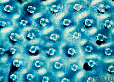
Agave Epidermis Lower Surface seen with Bright Field illumination |
Agave Epidermis Lower Surface seen with crossed Polarizers |
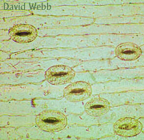
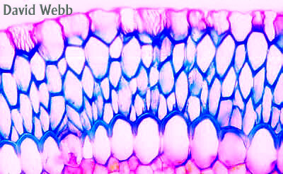
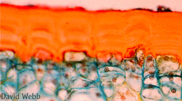
Uniseriate (Single Layer) Epidermis:
This is the most common type of
epidermis. 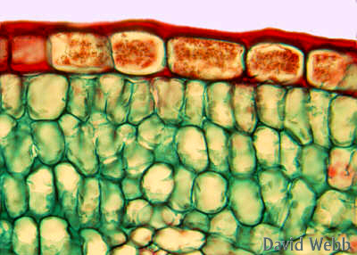
Examine commercial slides
The following features should be noted:
shape and size of epidermal cells
variation in size and structure of cells
cell contents.
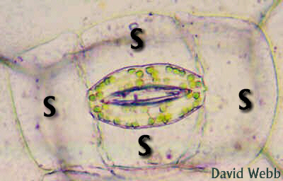
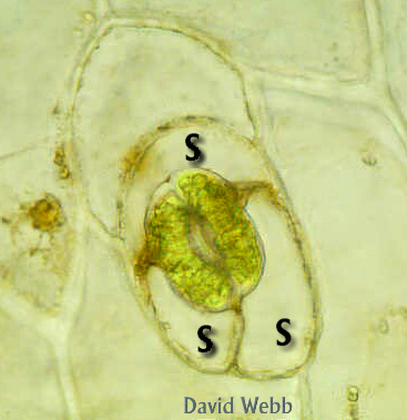
Note whether guard cells are in the same plane as rest of epidermis, or if they are raised or sunken.
Subsidiary Cells may or may not be present. The arrangement of subsidiary cells and guard cells can be used to identify plants.
We will study Pyrus (pear) leaves as an example of a
dicot.Sugarcane (Saccharum) or ko will be used as an example of a
monocot.A special feature to note is the prominent bulliform cells. These are involved in leaf expansion and in the folding of leaves subjected to drought.
Ancient Hawaiians brought many varieties of ko with them. The stems were sucked or eaten raw. Sugarcane eventually became a major plantation crop in Hawaii but this epoch is coming to an end.
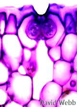
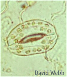
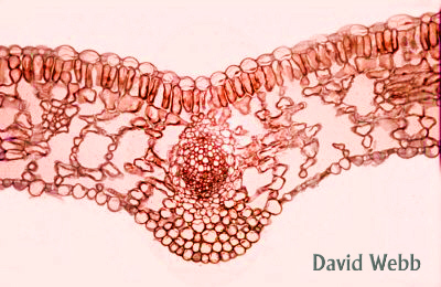
These images display various aspects of Epidermal anatomy with an emphasis on Stomata.
Agave Leaf stained with Toluidine Blue: Note the thick Cuticle which has stained light blue
Unstained Leaf Section: Locate the Guard Cells & decide if this is a Sunken Stomata or not. |
|
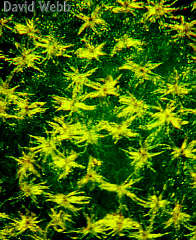
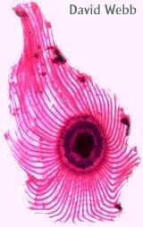
Stomata from the Hawaiian Silver Sword: How would you describe the surface position of these stomata???? |
|
|
|
|
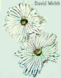
| Stomata without Subsidiary Cells | ||
|
|
|
| Two different Stomata with Subsidiary Cells | ||
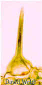
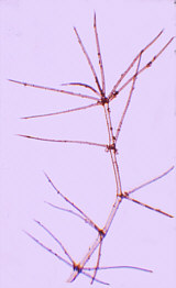
Examine whole mounts with a dissecting microscope, and the compound scope, and prepare proper sections for observation with higher magnifications on the microscope. Trichomes can be unicellular or multicellular; glandular or nonglandular.
|
|
Unicellular Glandular Trichome (Urtica) |
Multicellular Glandular Trichome (Coleus) |
Unicellular, multicellular and glandular hairs
Examine cross sections of Pentas or Widelia stems containing glandular trichomes.
Make cross sections of pubescent leaves from
|
|
Cross section of ohi'a lehua Leaf: Note the Trichomes (T) and the Cuticle (C) |
Same leaf viewed with crossed Polarizers |
Observe Dendroid hairs of Pterospermum and/or Candelabrum-like
 hairs on Verbascum
leaf.
hairs on Verbascum
leaf.
Stinging Hairs - Observe
commercial slides of Urtica, Observe fresh sections of Urtica
stem or leaves (if available).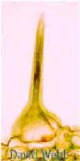 (Stinging Nettle) leaves.
Find the stinging hairs. These work like hypodermic syringes and inject a powerful
chemical upon contact.
(Stinging Nettle) leaves.
Find the stinging hairs. These work like hypodermic syringes and inject a powerful
chemical upon contact.
Closely related species in Hawaii have hairs that closely resemble their mainland cousins but lack the noxious secretion. This is probably due to the absence of herbivores in these islands. What is the advantage gained by NOT producing the secretion?
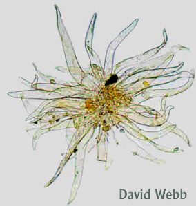
Scales or Peltate Hairs
are large complex structures which can resemble umbrellas or shields.
Add a drop of Toluidine Blue to the side of the coverslip and watch the wild staining reaction!!!!! Hawaiians called this plant ‘umi’umi-o-Dole or Dole’s Whiskers.
Similar Trichomes can be observed on Olive Leaves. These account for the Silver appearance of these leaves when they are dry.
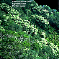
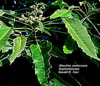
You may have noticed that kukui leaves are lighter than other leaves in the forest. This is due to the presence of multicellular nonglandular trichomes.
Observe
Scrape some onto a drop of water on a microscope slide add a cover slip and observe!!
|
|
| Kukui Trichomes | |
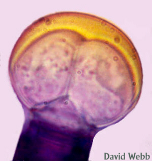
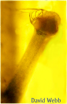
Large, complex Glandular trichomes are known as Colleters. These have multicellular stalks which can contain vascular tissue.
Study the Colleters on the calyx of Plumbago.
Use whole mounts and examine them with your
microscope by adding water & a cover slip.
|
|
|
Plumbago Bud |
Plumbago Flower |
Plumbago Colleter |
 Observe whole mounts and leaf sections of "Sundew (Drosera
sp.).
Observe whole mounts and leaf sections of "Sundew (Drosera
sp.).
Locate the complex multicellular glands (colleters) which trap insects.
Notice that the secretory portion of the colleter has vascular tissue in it.
What is the most probable source of the red pigment in Sundew Colleters?
There is a native Drosera (D. anglica)
|
|
|
Drosera Colleters |
||
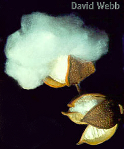
Cotton Fibers:
Observe DEMO slide of Gossypium (Cotton) fruit that shows young fibers which are epidermal hairs of the seeds.
Observe whole fruits and seeds if available.
Cotton is the most important vegetable fiber and has staged a commercial resurgence recently.
Hawaiian cotton has been useful in the genetic improvement of commercial cotton cultivars.
The presence of a multiple epidermis is rare and is restricted to the leaves of certain families like the Moraceae (Breadfruit & Figs) , and to orchid roots. The multiple layers can be traced back to the Protoderm which is the primary meristem for the Epidermis. Thus, two or more cell layers are derived from the protoderm. A good example of multiple epidermis is found in Ficus (Fig) leaves.
- Make free-hand cross sections of Ficus leaves. Note the many layers of achlorophyllous cells on the upper side of the leaf. These constitute a multiple epidermis. You would need to do a developmental study to be certain about this.
The epidermis of Ficus is also known for crystals called cystoliths which are found in certain epidermal cells called lithocysts.
Find these in the Ficus leaf sections and compare with commercial slides.
|
|
|
Ficus Leaf with a Multiple Epidermis & Lithocyst |
Section of an Orchid root with a Velamen |
Orchid Root: Mature Velamen covers the white area. |
The
- Make hand sections, Stain with Toluidine Blue and observe with and without polarizers.
Velamen stained with Toluidine Blue |
Velamen seen with crossed polarizers |
|
|
Olive Tree |
|
|
|