During this course we will examine each of these properties to some extent. For now we will content ourselves with just the first...cellular structure. All truly living things are composed of cells. These are microscopic chambers that each contain the "stuff" of life. In fact, the smallest organisms consist of just one cell! These unicellular organisms are found generally in the two Kingdoms called Monera and Protista. As organisms, these single cells carry out all of the properties of life listed above! Thus the cell is a unit of life.Properties of Life
cellular structure
homeostasis-metabolism
growth
reproduction
adaptation
short-term behavior
long-term evolution
It is important to realize, however, that in multicellular organisms such as higher plants and humans there are cells that lack certain properties. In fact some cells function best when they are totally dead as we shall soon see...
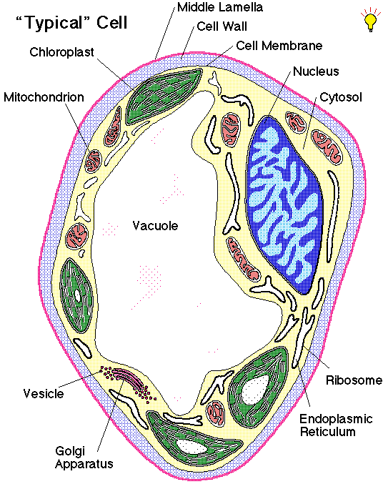
The cell shown below is called a parenchyma cell. It is perhaps typical of those found in the soft cells of all plants. A herbaceous plant (such as a grass) might be 90% parenchyma. A woody plant (such as a tree) might be less than 30% parenchyma.
As you can see in the diagram above, a plant cell has an outer boundary that includes a cell wall. This cell wall is unique to the range of organisms called plants...animal cells do not have cell walls! This wall is a boundary between the cell and its environment. Just like any good wall, it keeps out any invaders and other cells. But again, just like any good boundary, it is meant to be crossed. In fact the import and export of materials is critical to the life of the cell. So a wall is only as good as its doors and windows. This lesson was not well appreciated by Soviet states and ancient China. The exchange of materials and ideas provides vitality to what is within the wall. Without this exchange death is really the only option. The plant cell wall is quite passive really. It is made up of cellulose (an indigestible--by humans--polysaccharide carbohydrate) and other polymers of sugar. The fibrils of cellulose form a loose basket-weave arrangement through which materials can filter in or out without further barrier. It serves more of a filtration function than a control function. The other polymers in the wall area may include pectin (a polygalacturonan) that serves as the glue to hold one plant cell firmly to an adjacent cell. In fact the outer layer of higher plant cells is sometimes called the middle lamella because this heavy layer of pectin glue is quite visible in electron microscope images of plant tissues. Humans extract pectin and use it to thicken fruit juices into a material called jam or jelly (depending on the presence or absence of fruit pulp). You can buy Certo or Sure-Jell in the grocery store to use in jelly-making; these are purified forms of pectin--plant cell glue.
Just inside the cell wall is the cell membrane. This membrane is made up of phospholipids (such as lecithin) and membrane-proteins. This thin "skin" of the cell is the important import/export control area. The phospholipids provide the barrier functions. Their hydrophilic polar "heads" face the inside and outside of the membrane and prevent passage of hydrophobic substances through the membrane. The phospholipid hydrophobic "tails" face the middle of the membrane layer and prevent the passage of hydrophilic substances through the membrane. Thus the layered form of the membrane is a pretty good barrier to most chemicals. Special proteins made by the cell serve as windows and doors in this membrane. These transport proteins have both hydrophobic sections that align inside the membrane and hydrophilic sections that align with the inner and/or outer membrane surfaces. By bridging both the hydrophilic and hydrophobic regions of the membrane, these proteins are "bound" to the membrane. Most interestingly, however, is that these bridging proteins can form channels through the membrane for a particular kind of chemical to pass. In fact, some of the membrane proteins can energetically grasp chemicals and actively pump them across the membrane to the other side! These transport proteins are sometimes called active transport carriers. Thus membrane proteins are those all-important doors and windows through the barrier!
Moving to the middle of the cell somewhere we find a double membrane surrounding a region of fluid which contains DNA. This structure is called the nucleus. Please note the spelling and pronunciation (new-clee-us...NOT...new-kew-lus); learning correct pronunciation will help correct spelling (probably, library, and February are also commonly mispronounced and misspelled). Nothing makes you sound more ignorant of your studies than mispronunciation of its critical vocabulary. The nucleus processes information held in the DNA molecules that it contains. Sometimes the nucleus copies the DNA into new molecules of DNA in preparation for cell division; this process is called replication. Replication is a critical prelude to making two cells from just one and so is tied directly to reproduction and inheritance from one generation to the next. A more mundane function of the nucleus is to make an RNA copy of the DNA; this process is called transcription. The DNA is a more-or-less permanent copy of critical information; it might be thought of as master plans for the cell. When these master plans are to be used, they are transcribed into an inexpensive and disposable copy in RNA...a kind of blueprint to be handled, used, and ultimately destroyed. These RNA blueprints are sent out from the nucleus to be acted upon by other parts of the cell.
Just outside the nucleus and extending out to the cell membrane is an area often referred to as cytoplasm. We will avoid this all-encompassing term because it gives the idea that this part of the cell is somehow a uniform fluid. Indeed to ancient biologists using light microscopes this region did seem pretty homogenous--hence one word to describe it. With the advent of the electron microscope we have discovered other structures floating in this region of the cell and so we generally avoid the term cytoplasm. In its place we simply use cytosol to refer to the liquid around all of the floating structures between the nucleus and cell membrane. The cytosol is mostly water, but significantly contains many dissolved substances. Notable among these substances are enzymes. Enzymes are proteins that cause certain chemical reactions to occur at a much faster rate. These enzymes might build materials (anabolism) or take apart materials (catabolism) in the cell, depending upon the kind of enzyme in observation. Thus these enzymes are involved in the chemical processing that actively takes place in the cytosol. The whole of these processes is called metabolism; anabolism and catabolism are two opposing categories of metabolism. Thus much "work" is done in the cytosolic fluid of cells!
Some of the floating membranes in the cytosol include a network of tiny tubes that interconnect throughout the inside of the cell. This network is called endoplasmic reticulum...literally a network inside the fluid of the cell. This network is involved in the transport of materials from one part of the cell to another, and maintains the integrity of the membranes surrounding the nucleus. This is the conveyor belt of the cell.
On the surface of some of the endoplasmic reticulum are thousands of tiny structures in the cytosol called ribosomes. These ribosomes are made of protein and RNA and are actively involved in the synthesis of proteins. The ribosome attaches to the RNA blueprints coming out from the nucleus. It reads the information coded in the sequence of bases in the RNA and, using this information, assembles a particular protein. Ribosomes are thus a kind of protein-synthesis "machine." This process of using the information in RNA to make a protein is called translation; it is the complement of transcription. The protein products of the translation include the enzymes ("workers") of the cell. These proteins are shipped throughout the cell for functional uses.
The endoplasmic reticulum near the ribosomes is responsible for moving much of the protein through its tubules to other parts of the cell. One area of particular importance on the fringes of the endoplasmic reticulum is the Golgi apparatus or dictyosome. Here the proteins arrive and are sorted by tagging with sugars or lipids, and are packaged into tiny membrane-bound packages called vesicles. The Golgi apparatus thus is a sorting and packaging structure. Depending upon contents, these vesicles move to a particular destination and the membranes fuse. The contents of the vesicle are thus "dumped" to the other side of the receiving membrane.
This vesicular transport process explains how proteins made by the cell might be dumped through the cell membrane to the outside environment. Such a process is called exocytosis; literally a process pushing materials outside of the cell. On the other hand, vesicles can also be formed at the cell membrane by engulfing materials on the outside of the cell to bring to the inside. Such an import process would be called endocytosis if the material is particulate. In the special case of vesicular import of only water and dissolved chemicals, the process is named pinocytosis; literally cell drinking. Vesicular transport adds to the repertoire and to the scale of the import/export operations carried out at the cell membrane.
To drive almost all of the processes described above, we need energy. In animals there is one energy powerhouse...the mitochondrion. This structure has a smooth outer membrane and a convoluted inner membrane. The mitochondrion imports fuel molecules (chiefly acetyl Co-enzyme A) to be converted into carbon dioxide and water. This process requires the presence of oxygen and is called respiration:
CH2O + O2 --------> CO2 + H2O + energyYou might notice that this process is essentially the reverse of photosynthesis (though drastically different in details!) and releases energy instead of requiring the input of light energy. This release of energy from the respiration in the mitochondrion allows for all the work done in the cell. All living cells with a true nucleus (the eukaryotic cells), including those of plants, contain mitochondria and carry out respiration. Yes, again, plants have mitochondria and they do respiration both day and night! Many people are unaware of this fact...now you know!
It is easy to understand how the cell gets its oxygen from the atmosphere, but from where does it get the carbohydrate (CH2O)? In the case of fungi and animals, the fuels for respiration come from digestion of plant-derived foodstuffs found (or hunted -down!) in the environment. For plants, the carbohydrate can come from that essentially reverse process, photosynthesis:
CO2 + H2O + light -------> CH2O + O2Photosynthesis occurs in the chloroplast of the cell. This structure floating in the cytosol has two smooth outer membranes and a system of stacks of membranes inside. The whole chloroplast is some ten times larger than a mitochondrion, and it stacks of membranes inside are called grana. These grana are composed of many individual membrane sacs called thylakoid membranes. Each thylakoid membrane has special proteins that hold molecules of the green pigment chlorophyll. This chlorophyll is responsible for trapping the energy of the light to use in driving the production of carbohydrates in photosynthesis. We shall learn more of this process later in the term. For now, let's just remember that plants have both mitochondria and chloroplasts and therefore can do both respiration and photosynthesis! Animals lack chloroplasts.
Finally in our brief tour of cell structure and function, let us remember that all processes described here produce waste materials, and this leads us to one more distinction between plants and animals. Typically animals use various processes to dump their wastes to the environment. Single-celled animals may use vesicular transport to rid themselves of waste by exocytosis. Higher animals concentrate wastes through a urinary system and then urinate periodically in the environment. Since a plant produces great food for an animal to eat, you might not be surprised to find that plants hold their toxic wastes. This way a grazing animal gets a dose of poison every time it tries to eat the plant. The toxic wastes of a plant cell are stored in the vacuole. This central region is surrounded by a membrane (the tonoplast) that selectively pumps cellular wastes and poisons into the vacuole. Early botanists mistakenly thought the vacuole was pretty empty and uninteresting...hence the name vacuole...but now we know that this is a very interesting place where some pretty important metabolism occurs. Certain materials are recycled back out of the vacuole after detoxification reactions. Others are converted to even more-toxic forms once they reach the inside of the vacuole. Yet others are crystallized into raphides or other forms that pierce animal digestive systems or otherwise cause hemorrhage inside the herbivore. The vacuole is certainly not a vacuum nor void of interest!
There are many details left out of this brief description of cell structure and function, but at least we have made a start. One good way to think about the parts and functions of cells is to think of the cell as a business. How would you organize a factory that was making and exporting a particular product? A cell is well-designed to do this...
Another important way to study is to think about the three fundamental structures listed above that are unique to plants. Which ones are they?Cell Functions - The factory analogy
Cell Wall = border fence (very penetrable) good support
Cell Membrane = loading/unloading dock
Nucleus = administrative complex (DNA replication/transcription)
Cytosol = work floor environment
Endoplasmic Reticulum = conveyor belt system
Ribosomes = protein synthesis machine (translation)
Golgi Apparatus = sorting/packaging department
Vesicles = import/export package
Chloroplast = energy source (photosynthesis)
Mitochondrion = energy source (respiration)
Vacuole = toxic waste pond (herbivore poison)
Organelles unique to plants:
- Cell Wall
- Chloroplast
- Vacuole
Note: the above are in addition to the usual organelles found in animal cells. Plants are MORE...not less....than animal cells!
Finally, a good way to study cell structure and function is by means of a concept map. Here is a crude concept map showing the relationships between the parts in terms of functions.

I mentioned above that the smallest plants consisted of single cells. The largest organism on our planet is the sequoia tree. This tree is larger than 10 blue whales (the largest animal) and so is easily the largest obvious organism. Certain fungi in soil, however, consist of microscopic but connect filaments of cells originating from a single spore. These fungi are basically invisible, but DNA evidence suggests that single individuals may spread across hundred of acres of land. So these fungi may, in fact, be the "largest" organisms.
In terms of lifespan, some cells (and unicellular organisms) live for only a few minutes before dividing and making new cells or perishing altogether. Some multicellular organisms can live to be very old. The oldest documentable living organism is the bristlecone pine tree. These trees are frequently more than 5000 years old. They were already older than any extant human when the pyramids were built in Egypt! These plants, still alive today, were already 3000 years old when Jesus walked through Jerusalem. Admittedly, some clonal plants are harder to document and may be even older than the bristlecone pine, but the age of the bristlecone pine is easy to document!
You might be misled into thinking that the "typical" cell is most of what is available or most of what is important. This couldn't be farther from the truth! Plant cells can be classified into four fundamental cell types (and many other specific kinds).
Meristematic Cells undergo mitosis and thereby produce all cells of the plant body. Mitosis is the nuclear division (usually followed by cytokinesis) that results in the production of new cells. Mitosis is a process in which a cell goes from interphase, through prophase, metaphase, anaphase, and telophase, and ends up in interphase again. The cell division process assures that the master plans in the DNA of the nucleus are replicated accurately and completely, and that the copies of the master plans are evenly divided between the two resulting cells in an orderly fashion. This ensures the integrity and completeness of the master plans in both of the cells.
Derivatives of meristematic cells can mature in any one of three directions...only one of which results in a "typical" cell (parenchyma):
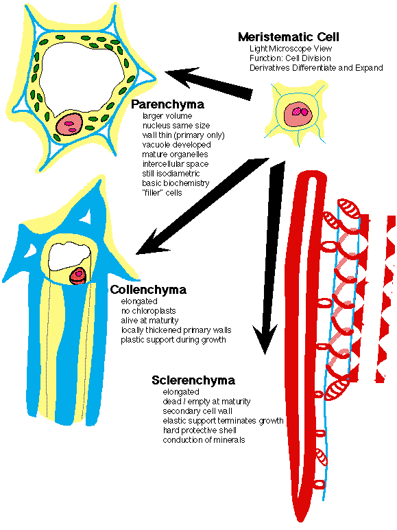
It might not be completely obvious, but study of the diagram above will reveal that some cells are long and thin, some cells have thin walls and others have thick walls, and some cells are alive and others are dead!
Indeed dead cells can be useful to a multicellular organism. In humans, dead cells contribute the structure of hair shafts and nails. In plants dead cells provide stony protective layers in peach pits and the conductive elements of the xylem. Without the dead xylem elements, a plant would literally cook in the sun because it would lack the water from the xylem to efficiently cool the leaves by evaporation of that water. Without the dead xylem to bring minerals up with that water, plants would lack essential metal ions from the soil. These soil minerals include calcium, magnesium, iron, and zinc (more on those later in the course!). Our food would lack some of its essential nutrition were it not for these dead cells in plants!
This page © Ross E. Koning 1994.
The MLA citation style for this page would be:
Koning, Ross E. "The Unit of Life". Plant Physiology Website. 1994. http://koning.ecsu.ctstateu.edu/plants_human/unitlife.html (your visit date).
Go back to the Course Schedule.
Go back to Ross Koning's Home Page.
Send comments and bug reports to Ross Koning at koning@ecsu.ctstateu.edu.
View the Standard Disclaimer.