Botany online 1996-2004. No further update, only historical document of botanical science!
Proteins as Analytical Devices
(Probes) for the Localization of Molecules and for the Research into
the Molecular Architecture of Cells
Since the second part of the previous century, dyes are used to
detect certain classes of compounds in microscopy. During the
thirties of this century, fluorescent dyes that could be used in much
lower concentrations as 'vital dyes' became popular.
Colour molecules are usually small and their disadvantage is
therefore a rather poor specificity. There exist, for example,
protein-specific dyes but hardly any that are specific for a certain
enzyme. A selective binding could be shown to exist for some toxins
(fungi toxins, for example). Phalloidin (toxin of the amanita) binds
to actin. By coupling a fluorescent dye to phalloidin, actin can thus
be localized within the cell .
The different macromolecules and especially the proteins are far
more specific:
Lectins. We got to know lectins in the
section before last. Each lectin can be coupled to a fluorescent dye
like fluoroisothiocyanat (FITC: green fluorescence) or rhodamine (red
fluorescence). Such preparations are used in a big way in medicinal
research and are on the market at rather low prices. They are
well-suited (as we will often see in pictures presented in
Botany online) as probes
for the localization of glycoconjugates at
cell surfaces, membrane surfaces, at compartments and others.
Macromolecules cannot easily penetrate cells. They are therefore
primarily markers for extracellular surface receptors. By treatment
with cellulase, for example, the cell wall of plants can be degraded,
the result are protoplasts without cell
walls. Depending on their origin, they bind ConA or RCA indicating
that glucosyl-or mannosyl residues resp. galactosyl-residues are
present at their surfaces. Protoplast preparations contain often a
whole range of different cell fragments, among others also free
vacuoles. The vacuolar membrane, the tonoplast, binds to no known
lectin. This alone shows that it is not organized like the plasma
membrane.
Fluorescence tagged lectins are especially well-suited for the
mapping of lectin receptors in or at cell walls, for the proof of the
cell's polarity, to detect different states of cell activity or
certain states of the cell cycle; they are suited for the solubility
properties of lectin receptors. WGA is an indicator
for fungi mycelin in infected plant tissue.
For electron microscopic studies, the lectins have to be tagged
with electron-dense markers like ferritin or colloidal gold. These
complexes enable the detection of different distribution at the
membrane's inner and outer surface. Lectins have also some
disadvantages:
They bind to so-called glycoconjugates. It can therefore not
be said without further analysis whether the receptor is an oligo-
or a polysaccharide, a glycoprotein or a glycolipid.
Lectins detect only a rather small range of sugars. There
exist no lectins that detect, for example, arabinose or xylose
residues.
 Antibodies. Antibodies are a rather homogeneous
group of proteins that occur in the serum of vertebrates protecting
the animal from foreign influences like infections with bacteria or
viruses or from tumour cells. Plants have no antibodies. But since we
need them for the study of plant cells, will we outline them briefly:
Antibodies. Antibodies are a rather homogeneous
group of proteins that occur in the serum of vertebrates protecting
the animal from foreign influences like infections with bacteria or
viruses or from tumour cells. Plants have no antibodies. But since we
need them for the study of plant cells, will we outline them briefly:
Antibody production can be induced (immunization), i.e. a signal
is needed that stimulates the animal organism to generate antibodies.
Such a signal has to be of a macromolecular nature. It may be part of
a cell surface. A component that induces antibody production is
called antigen. An antigen is
usually bigger than the respective binding site of the antibody. The
antibody-binding domain of the antigen is called an
antigen determinant. At an
immunization, as many different antibodies are produced as antigen
determinants are present. An antibody population is thus always
heterogeneous or, as it is called,
polyclonal.
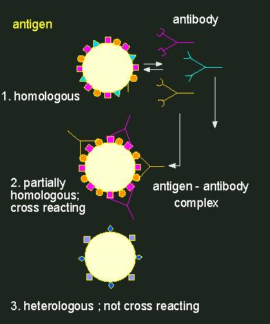
Antigen-antibody reaction.
Antibodies (depicted as Y-shaped structures) form a
heterogeneous population of molecules with different
specificities. A cross-reaction of an antibody population (an
anti-serum) with a foreign antigen (in the middle) occurs only, if
the homologous and the foreign antigen are at least partially
equipped with the same determinants. Every antibody has two
identical binding sites for antigen determinants.
Normally, rabbits are used for the production of antibodies with a
certain specificity. Two to three weeks after immunization, some of
their blood can be extracted. After centrifuging the blood cells
down, an antiserum is gained that contains the specific antibodies.
They can now be used for the qualitative or quantitative detection of
the used antigen or for the detection of substances that are similar
to the antigen. In such cases it is spoken of serological
cross-reactions or serological relationships. A more or less clear
serological relationship is usually found in homologous proteins
(enzymes, storage proteins, cytochrome C, etc.) that were gained from
more or less closely related animal or plant species. The degree of
serological relationship is normally related to the degree of
different amino acids in the
proteins.
A number of partly rather sensitive serological tests, like for
example the radio immuno assay (RIA)
exist.
If an antibody against a rather small molecule like a phytohormone
is needed, the antigen has at first to be coupled to a larger
molecule. It does thus gain the property of an antigen determinant
and among the numerous antibodies produced will also be some that are
directed against the phytohormone. They can easily be separated from
the other antibodies that do all react with the coupled larger
molecule by precipitation. The remaining antibodies will all be
directed against the hormone.
To localize antigens in cells, the method that has already been
described for lectins is chosen. But it is not common to tag an
antibody directly with a fluorescent dye, since it is much more
comfortable to produce antibodies against rabbit antibodies in
another animal (for example in goats), to gain these in larger
amounts and to tag them with the fluorescent dye. This is termed
indirect fluorescence. Why this
detour?
- Goat-anti-rabbit-antibodies can be used against every antibody
generated in rabbits.
- The specific rabbit-antibodies are normally only available in
small amounts. Often, a number of rabbits are immunized in
parallel against different antigens. Each of these different
antibodies would have to be tagged with the fluorescent dye.
- The use of a second antibody amplifies the fluorescence since
several goat antibodies are bound by one rabbit antibody.
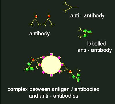
Indirect immunofluorescence.
1. Specific antibody
against the primary antigen. The antibodies themselves have
antigen determinants (indicated by red circles). Normally,
antibodies of this kind are generated in rabbits.
2. Anti-antibody (generated,
for example, by the immunization of a goat with rabbit
antibodies). These anti-antibodies (goat against rabbit) bind to
the antigen determinants of the rabbit antibodies. If they are
labelled with a fluorescent tag (green)
(3), a fluorescent complex at
the primary antigen is the result
(4).
For electron microscopic methods again, antibodies are tagged with
electron-dense materials.
However broad the range of uses in medicine, in plant research
antibodies are still used only rarely. One of the main reasons is the
cell wall that cannot be penetrated by any antibody, the other reason
is the lack of well-suited antigens. But still, several good results
do exist. Phytochrome, a light receptor that is among the
most-important control units in the plant could be localized
in certain cells. It could also be shown that it is missing in
others.
To mark cell contents, the cells have either to be cut into slices
or protoplasts with partially permeable membranes have to be
generated. Among the other antigens localized by immunofluorescence
are some enzymes like phosphoenol pyruvatcarboxylase,
alpha-amylase and several storage proteins and cytosceleton
elements.
Since their detection by C. MILSTEIN and G. KÖHLER (Medicinal
Research Council, Laboratory of Molecular Biology, Cambridge and
Basel Institute of Immunology) in 1975,
monoclonal antibodies are
regarded as the non plus ultra. Monoclonal antibodies are
homogeneous antibody populations that are produced in cell cultures
and that display a very narrow specificity (against only one
determinant). About their production:
Antibodies are generated in small lymphocytes. One of the tissues
with most lymphocytes is the spleen. Several days after immunizing,
for example, a mouse is its spleen removed and a cell suspension is
generated. These cells are then fused with myeloma cells of mice (one
of many tumour lines). The fusion produces hybrid cells (hybridoms).
Myeloma cells are characterized by unlimited growth. This property is
also kept by the hybridoma cells. But they have the additional
property of antibody production and secretion. In an intermediate
step is a wanted cell line isolated and cultivated.
The method has mainly one disadvantage: the antibodies are highly
specific but the number of binding sites (antigens) of the cells is
drastically reduced. Consequently, only very few antibody molecules
are bound, often is the antigen-antibody reaction at the limit of
provability. But here, too, exist ways to success: in indirect
fluorescence, TV cameras with electronic image intensification are
used.
Detection of Enzyme
Activities. Can enzymes or substrates in the cell also be
detected without antibodies?
Different methods exist. Most common is autoradiography. For this,
a radioactively tagged substrate or, even better, a substrate
analogue that is bound by the enzyme but not converted is needed.
After washing off the material that has not been bound, an
autoradiogram can be produced with the help of a special film that
shows the position of the enzyme. The method is especially well
established with membrane bound enzymes.
Another possibility is the use of a substrate that is processed
and renders an insoluble, stained product. In electron microscopy
again, electron dense markers are used.
© Peter v. Sengbusch - Impressum