Botany online 1996-2004. No further update, only historical document of botanical science!

©
Rafael Tormo Molina
|
Karyokinesis and Cytokinesis - Mitosis
In 1888, the longitudinal structures that could
be observed with the microscope during mitosis were named
chromosomes. The
state at the beginning of mitosis, when they become
visible was termed
prophase. After the
prophase the chromosomes arrange at the equatorial plane,
a state called
metaphase.
Subsequently the halves of the chromosomes, the
chromatids, are torn
to the two opposing cell poles. This phase is called
anaphase. Finally,
during telophase, the
two daughter nuclei form.
While the chromosomes condense, the spindle fibres are
assembled within the plasma. They form a structure called
the mitotic spindle
that provides the means for the separation of the
chromatides. It consists of two different types of
fibres, the polar
microtubules that stretch from pole to pole
and the kinetochore
microtubules, that connect pole and a certain
area of the chromosome, the kinetochore. As the name says
the fibres are composed of microtubules. Their subunit is
tubuline.
|
Normally cytokinesis and karyokinesis (divisions of cell and
nucleus) are coupled. Exceptions do exist, but they will be discussed
elsewhere. The process of the division of
the nucleus in plant cells was elucidated by E.
STRASBURGER and published in his fundamental work "Zellbildung
und Zellteilung" (Cell Formation And Cell Division) in 1875. Already
in 1884 the results had been part of his 'Kleines botanisches
Praktikum' (A Short Course on Botany) and since 1894 they were an
established part of his "Lehrbuch der Botanik" (A Textbook on
Botany). In the 4th edition the division of the nucleus is described
as follows:
" Except for special and very limited cases plant cell nuclei
propagate by the so-called mitotic or indirect division. The
process is also called karyokinesis (today usually mitosis). It is
a rather complicated process that seems to be necessary to
distribute the substance of the mother nucleus evenly on both
daughter nuclei."
At first STRASBURGER did work with material that had been fixed
with alcohol. But in 1879 A. LUNDSTRÖM was able to investigate
the mitosis of a living specimen, the anthers of Tradescantia.
Modern microscopic techniques, especially phase contrast and
interference contrast microscopy allow today to make the process
of the division of the nucleus visible in a lot of cell
types.
Impressing educational films for schools on this process exist, so
that every student of biology should actually be familiar with it
even before the beginning of his study.
|
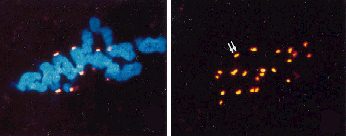
Confocal laser scanning micrograph of an anaphase onion
root tip cell showing immunocytochemical labelling of the
Golgi apparatus and plasma membrane
©
C. HAWES.
|
STRASBURGER's possibilities were much humbler. Even though he did
notice that the nuclei stretch before or at the beginning of mitosis
and take on a spindly shape. Longitudinal structures become visible.
He saw states, where they shortened and finally appeared as compact
little rods. In 1888 they were termed
chromosomes (Greek:
chroma=colour; soma=body) by WALDEYER, because they
were particularly well stained with a certain nuclear dye.
The state at the beginning of mitosis, where the chromosomes
become visible was termed
prophase after a suggestion of
STRASBURGER. The German anatomist W. FLEMMING recognized in 1880 that
the chromosomes of the prophase are characterized by a longitudinal
gap.
After the prophase the chromosomes are arranged
at the plane of cell division forming the
equatorial plane. This state is
called the metaphase. Both chromosome
halves (the chromatids) are
clearly recognizable. Consequently they separate in opposite
directions to form the two daughter nuclei. The phase of separation
is termed anaphase, the formation of the
daughter nuclei telophase.
Other processes are also involved. While the chromatin becomes
shorter, disentangles and separates into single chromosomes, fibrous
looking aggregates form in the plasma that strech from pole to pole.
They are called spindle fibres
and the whole structure has the name mitotic
spindle. The spindle is composed of undisrupted fibres
(they are bundles or aggregates of overlapping microtubules)
that stretch from pole to pole (polar
microtubules) and of other fibres that connect the pole
with a chromosome (kinetochore
microtubules). The separation of the chromatids is caused
by the contraction of the kinetochore microtubules. The kinetochore
microtubule is attached to a certain area of the chromosome, the
kinetochore, that each chromosome
develops during late prophase . Depending on its location the typical
V-, L-, U- or I-shaped structures of the anaphase form.
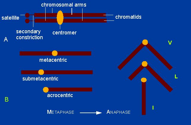
It is known today that the fibres contain tubuline,
the subunit of microtubules. It can be stained selectively with
fluorescence-tagged antibodies.
The spindle fibres are in fact bundles of
microtubules. During late anaphase the new cell wall becomes visible
in the equatorial plane. The wall
formation is completed during telophase.
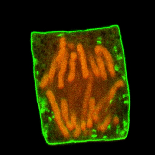
In contrast animal cells divide by constriction of the mother
cell. The decisive importance of mitosis in both cases is the
qualitatively and quantitatively exact distribution of the hereditary
material, whereby the longitudinal splaying represents the most
important point. Each daughter cell does accordingly inherit one half
of a chromosome, a chromatid. The
number of chromosomes is usually specific for a certain species. This
discovery was also made by E. STRASBURGER and the zoologist RABL from
Prague showed that it applies to animal cells, too. Exceptions are
caused, when single chromosomes stick together at their ends, whereby
the number of chromosomes changes. Such changes together with the
simultaneous genetic changes proved finally that the theory
of chromosomes was right.
© Peter v. Sengbusch - Impressum