Botany online 1996-2004. No further update, only historical document of botanical science!
All eucaryotic cells are interspersed with extensive membranous systems that structure the cell's interior into numerous compartments. The membranes are of uniform thickness and consist of a lipid bilayer. Among the membranous systems of the cell are the plasmalemma or plasmamembrane, the Golgi apparatus and the endoplasmatic reticulum (ER). The ER forms a continuous system with the nuclear envelope. The latter is composed of two lipid bilayers perforated by the nuclear pores.
Organelles are membrane-bordered functional unities of the cell. Both mitochondria and plastids have an inner and an outer membrane. Plastids have an additional internal membrane system, the thylakoid system. Mitochondria occur in animal as well as in plant cells, plastids only in plants. Microbodies are surrounded by only one membrane. They harbour specific metabolic pathways and are, dependent on these, classified as peroxysomes or glyoxysomes.
The picture (above) shows an electron microscopic image of a 'typical' plant cell at low resolution. It depicts several structures that we know by now: cell wall, nucleus, chloroplasts, vacuole and a thin rim of cytoplasm right at the cell wall. The picture does also show clearly recognizable substructures.
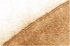 |
The most important insight of the first phase of electron microscopical research (from 1945 till about 1955) was that the ultrastructure of animal and plant cells is nearly identical. The cells are interspersed with extensive membranous systems that structure the plasma into a number of compartments. All membranes are of equal thickness and can be identified as double layers (two dark lines in parallel - see picture to the right). This is due to the uptake of the contrasting agent by both the in- and outside of the membrane. Based on their position within the cell and their association with other structures can membranes of different organelles or structures be classified : |
| |
The plasma membrane (plasmalemma) surrounds every cell |
| |
The endoplasmatic reticulum (ER) is an extensive intracellular membrane system, typical for all eucaryotic cells. Often are clearly defined particles, the ribosomes, found at the cytoplasmic side of the ER. These are the sites of protein synthesis. It is distinguished between rough (with ribosomes) and smooth (without ribosomes) ER, since not all ER bears ribosomes. |
| |
| |
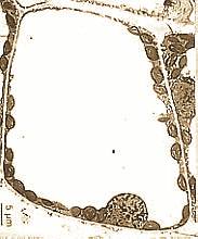
The nuclear envelope and the nuclear pores. The nucleus is enclosed by a double membrane, the nuclear envelope. The inner and the outer nuclear membrane are in contact with each other at special sites, the pores. They are not merely holes in the membrane, but rather a kind of very short tunnel, whose inner walls are a continuum of inner and outer membrane. Each pore has a complicated structure, called the pore complex. The pores provide a direct passage for the exchange of material between nucleus and cytoplasm. Numerous pictures show that the endoplasmatic reticulum and the outer nuclear membrane are a continuous membranous system. |
Nuclear envelope with nuclear pores. (Redrawn from W. R. BOWEN, 1969)
| |
The Golgi apparatus constitutes an additional membrane system of the
cell. The term refers collectively to all dictyosomes of a cell. Dictyosomes
are groups of flat, disc-shaped, vesiculous sacs or cisterns, often located
in vicinity to the ER. Dictyosomes of higher plant cells have usually four
to eight cisterns stacked together. These sacs do normally branch into
series of tubules and free vesicles at their margins. The Golgi apparatus
is an important coordination point of the cell's vesicular flow and of
its molecular export.
|
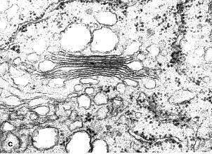  | |
(Ch.GLOCKMANN, R. KOLLMANN, 1984) On the right: Dictyosomes and dictyosomal vesicles (E. SCHNEPF) |
Organelles are membrane-surrounded functional unities of the cell. Mitochondria, the sites of respiration, and plastids, of which chloroplasts are the most striking members are maybe their best-known representatives.
| |
Mitochondria occur both in animal and plant cells, plastids only in the latter. Mitochondria consist of two membranes, the inner and the outer membrane, who form no continuous system. The inner membrane is strongly folded with the folds expanding into the inner of the mitochondrion, the matrix. Depending on the type of fold are mitochondria of cristae- and tubuli-type distinguished. |
| |
The plastids contain three different types of membrane. The inner and the outer membranes of the plastids form the plastid's envelope. Within this envelope lies with chloroplasts the thylakoid system that can be recognized in fully developed chloroplasts by a specific folding pattern. It occurs in other plastids, proplastids or different developmental stages of the chloroplast in simplified versions. The chloroplasts of algae are structured differently. Amyloplasts are starch-containing plastids. |
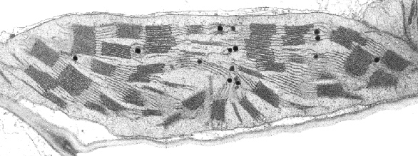
Thin section through a chloroplast of barley. Shape and arrangement of the grana are typical for normal chloroplasts. The dark circles are fat droplets (inclusions). Grana (stacks of thyacoids) may contain up to 25 thylacoids. (K.R. MILLER, Harvard University, 1976) | |
| |
Microbodies are cellular compartments of about half the size of a mitochondrion, which are surrounded by a simple membrane. Very specialized metabolic pathways take place within them. Depending on the type of chemical reactions is it distinguished between peroxysomes and glyoxysomes. |
| |
The nucleus, enclosed by the nuclear envelope, contains a granular looking material, the chromatin. The nucleolus can be seen as an area of high contrast. Further details became known after partial isolation and electron microscopical analysis of chromatin parts. |